File:Trichomonas.jpg
Trichomonas.jpg (580 × 435 pixels, file size: 142 KB, MIME type: image/jpeg)
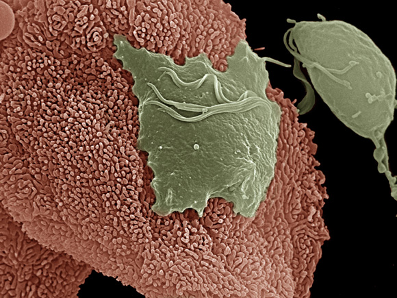
"An electron micrograph depicts the Trichomonas vaginalis parasite adhering to vaginal epithelial cells collected from vaginal swabs. A non-adhered parasite (right) is pear-shaped, whereas the attached parasite is flat and amoeboid." http://images.google.com/imgres?imgurl=http://www3.niaid.nih.gov/NR/rdonlyres/58C1A3DB-3E32-4DB1-8745-12D934001AA5/0/Tvaginalisparasite.jpg&imgrefurl=http://www3.niaid.nih.gov/news/newsreleases/2007/TvaginalisParasite_photo.htm&usg=__gzX94eZXxqu9s6igyPNcrvPdEaA=&h=435&w=580&sz=143&hl=en&start=5&sig2=1oBJxUYMe6zFAwTnUUiSfw&um=1&tbnid=Qk2CclPxMn2gsM:&tbnh=101&tbnw=134&prev=/images%3Fq%3Dtrichomonas%2Bvaginalis%26hl%3Den%26client%3Dfirefox-a%26rls%3Dorg.mozilla:en-US:official%26sa%3DN%26um%3D1&ei=Q4KVSvDNGouCNJ6gqPsH
File history
Click on a date/time to view the file as it appeared at that time.
| Date/Time | Thumbnail | Dimensions | User | Comment | |
|---|---|---|---|---|---|
| current | 18:58, 26 August 2009 | | 580 × 435 (142 KB) | Mlouden (talk | contribs) | "An electron micrograph depicts the Trichomonas vaginalis parasite adhering to vaginal epithelial cells collected from vaginal swabs. A non-adhered parasite (right) is pear-shaped, whereas the attached parasite is flat and amoeboid." http://images.google. |
You cannot overwrite this file.
File usage
The following page uses this file:
