File list
From MicrobeWiki, the student-edited microbiology resource
This special page shows all uploaded files.
| Date | Name | Thumbnail | Size | User | Description | Versions |
|---|---|---|---|---|---|---|
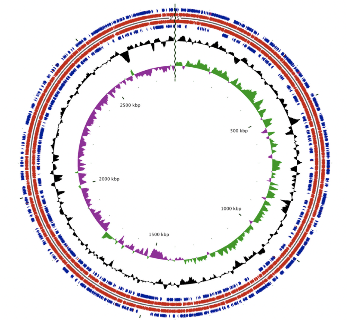
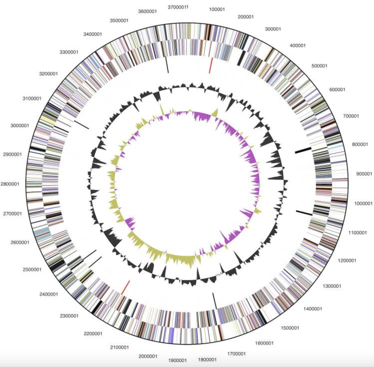
| 23:23, 24 April 2024 | Albertensis genome.png (file) |  |
1.3 MB | Raschnab | 1 | |
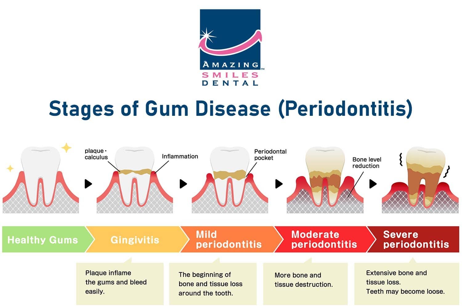
| 19:50, 24 April 2024 | Stages-of-Gum-Disease-Periodontitis.jpg (file) |  |
93 KB | Hrpeders | 1 | |
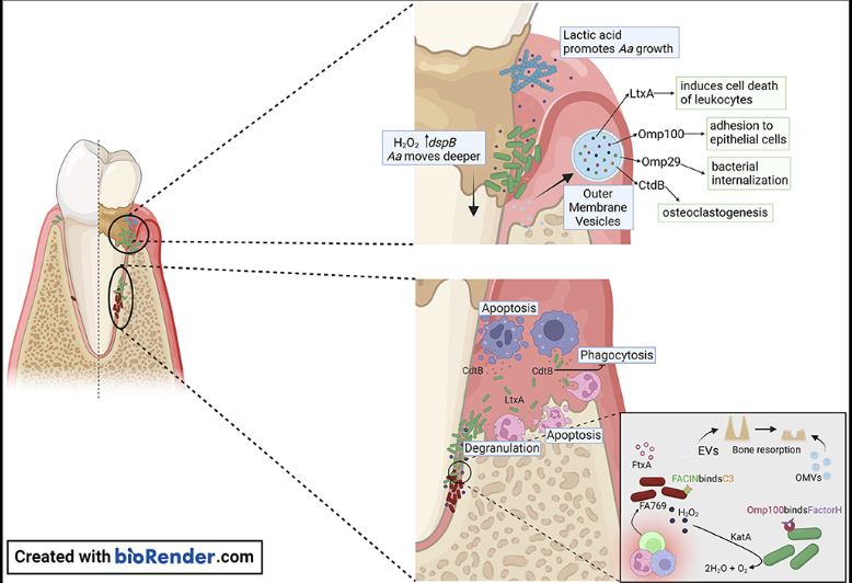
| 19:40, 24 April 2024 | Screenshot 2024-04-24 at 3.38.28 PM.png (file) |  |
370 KB | Hrpeders | 1 | |
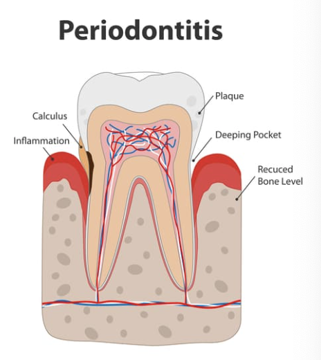
| 19:11, 24 April 2024 | Screenshot 2024-04-24 at 3.10.59 PM.png (file) |  |
231 KB | Hrpeders | 1 | |
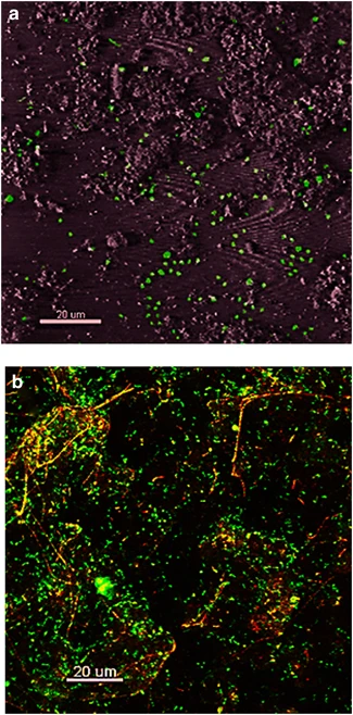
| 15:17, 23 April 2024 | 41396 2013 Article BFismej2012132 Fig2 HTML.webp (file) |  |
89 KB | Antieman | Confocal Microscopy of Fe-oxide microbial mat. | 1 |
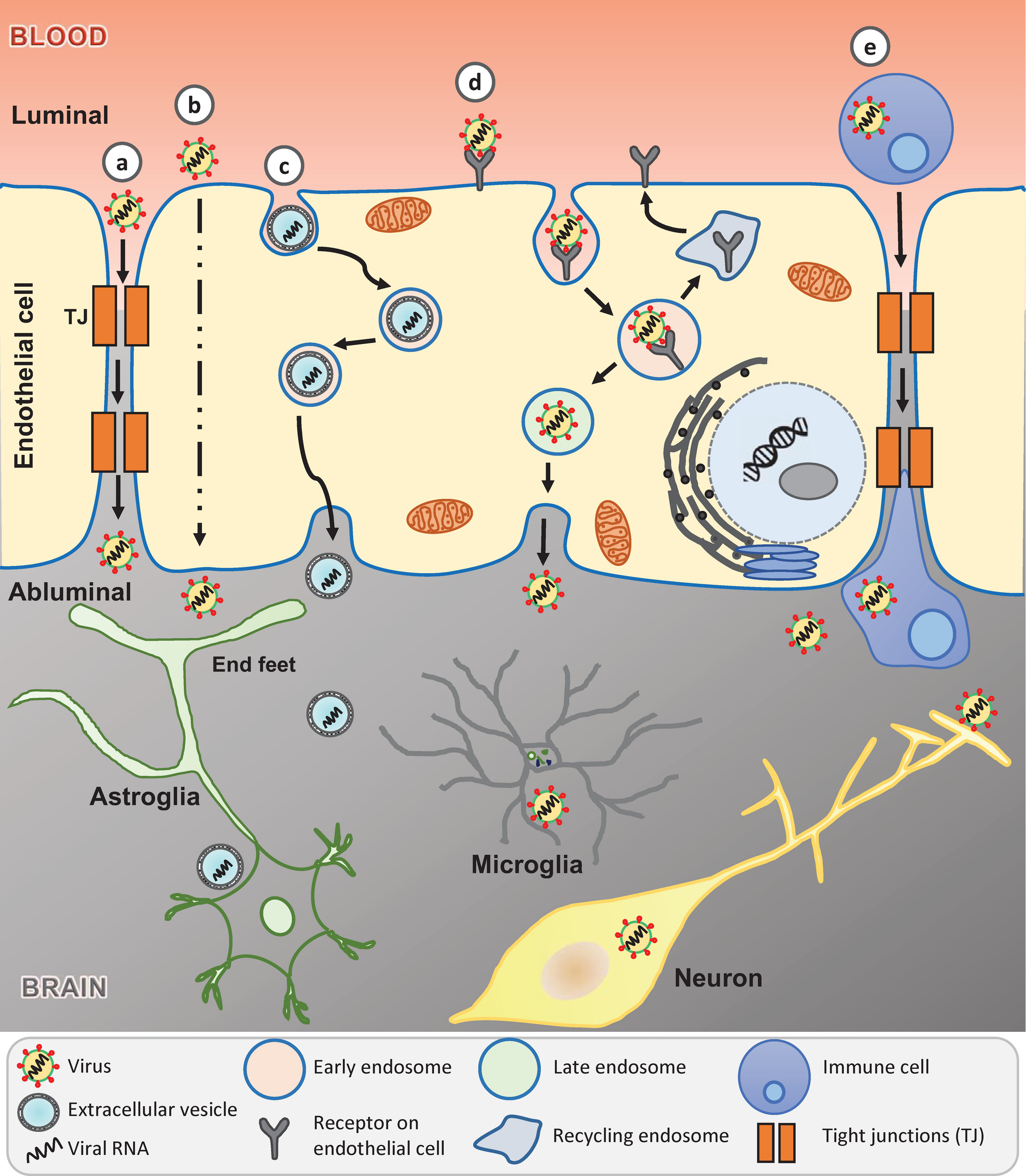
| 21:30, 22 April 2024 | Astroinfection.jpg (file) |  |
136 KB | Ehaggin | 1 | |
| 20:39, 22 April 2024 | Astrostructure.jpg (file) |  |
226 KB | Ehaggin | 1 | |
| 01:39, 18 April 2024 | Gut-Brain-Axis-1.jpeg (file) |  |
60 KB | Chlupsa1 | 1 | |
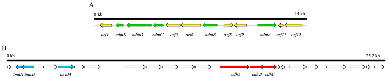
| 01:28, 18 April 2024 | Gutgeneex.jpeg (file) | 21 KB | Chlupsa1 | 1 | ||
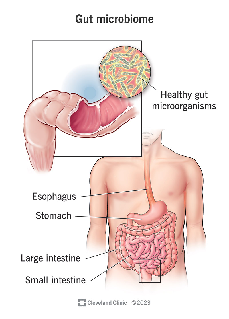
| 01:25, 18 April 2024 | Gutmicrobiome1.jpeg (file) |  |
125 KB | Chlupsa1 | 1 | |
| 14:05, 17 April 2024 | Screen Shot 2024-04-17 at 9.31.54 AM.png (file) |  |
1.75 MB | Vbpowell | 1 | |
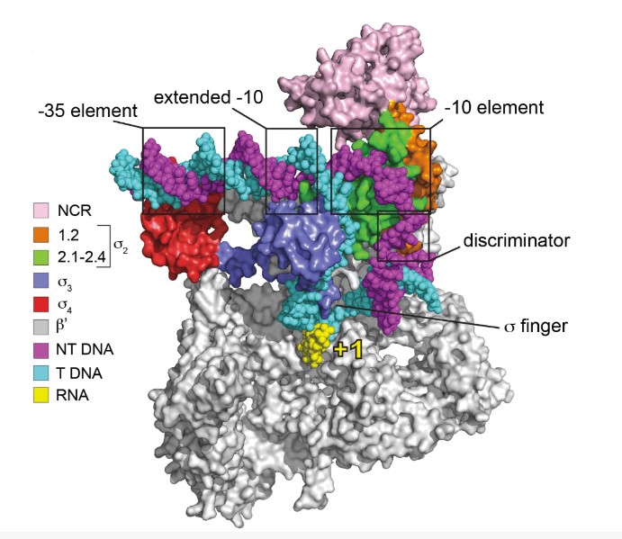
| 01:17, 17 April 2024 | Sigma.jpg (file) |  |
94 KB | Pardue | 1 | |
| 00:22, 17 April 2024 | Nitrite.jpg (file) |  |
28 KB | Pardue | 1 | |
| 04:48, 16 April 2024 | Evoh copolymer.png (file) |  |
3 KB | Pardue | 1 | |
| 18:04, 15 April 2024 | Orthhantavirus.jpeg (file) |  |
424 KB | Melo1 | 1 | |
| 18:03, 15 April 2024 | Hantavirus TEM.jpeg (file) |  |
187 KB | Melo1 | 1 | |
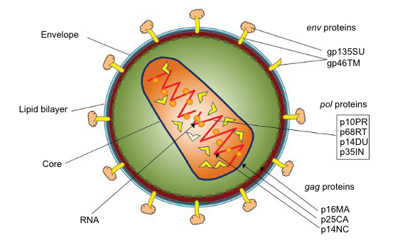
| 06:32, 15 April 2024 | SRLV particle.png (file) |  |
201 KB | Caine1 | 1 | |
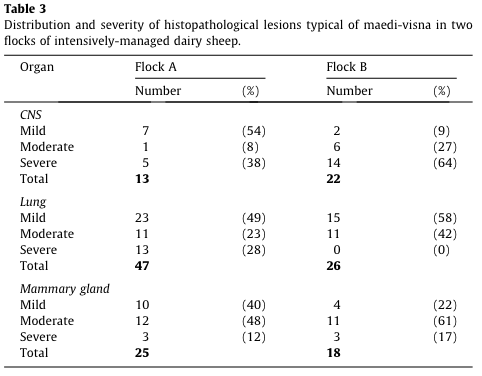
| 06:04, 15 April 2024 | Meadi-Visna lesions.png (file) |  |
45 KB | Caine1 | 1 | |
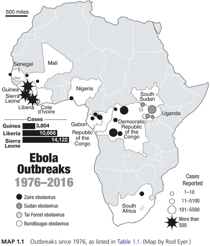
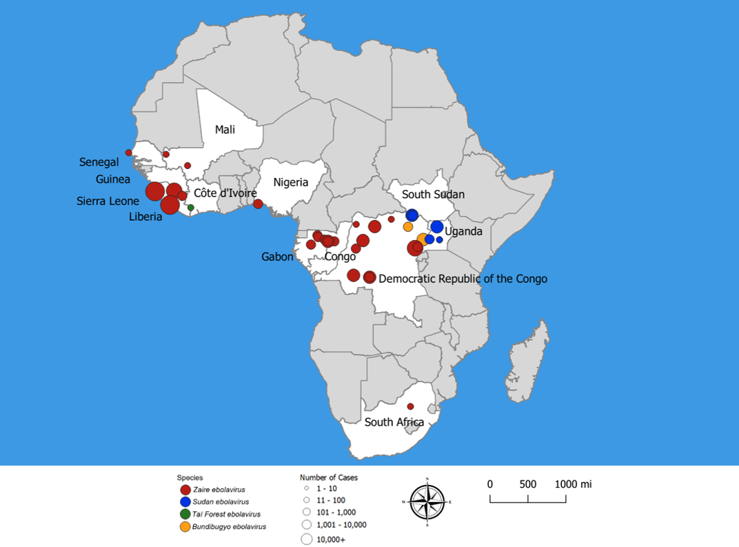
| 03:18, 15 April 2024 | EbolaOutbreaks.png (file) |  |
304 KB | Breard1 | 1 | |
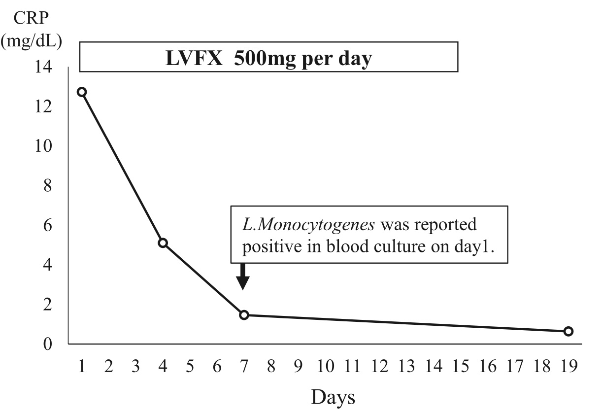
| 02:05, 15 April 2024 | CRP during antibiotic therapy.jpeg (file) |  |
96 KB | Chu2 | 1 | |
| 02:02, 15 April 2024 | Creep.png (file) |  |
904 KB | Leclerc1 | 1 | |
| 02:00, 15 April 2024 | Direct.jpeg (file) |  |
740 KB | Yang | 1 | |
| 01:59, 15 April 2024 | Skinwalker.png (file) |  |
920 KB | Leclerc1 | 1 | |
| 01:56, 15 April 2024 | Color3.jpg (file) |  |
296 KB | Leclerc1 | 1 | |
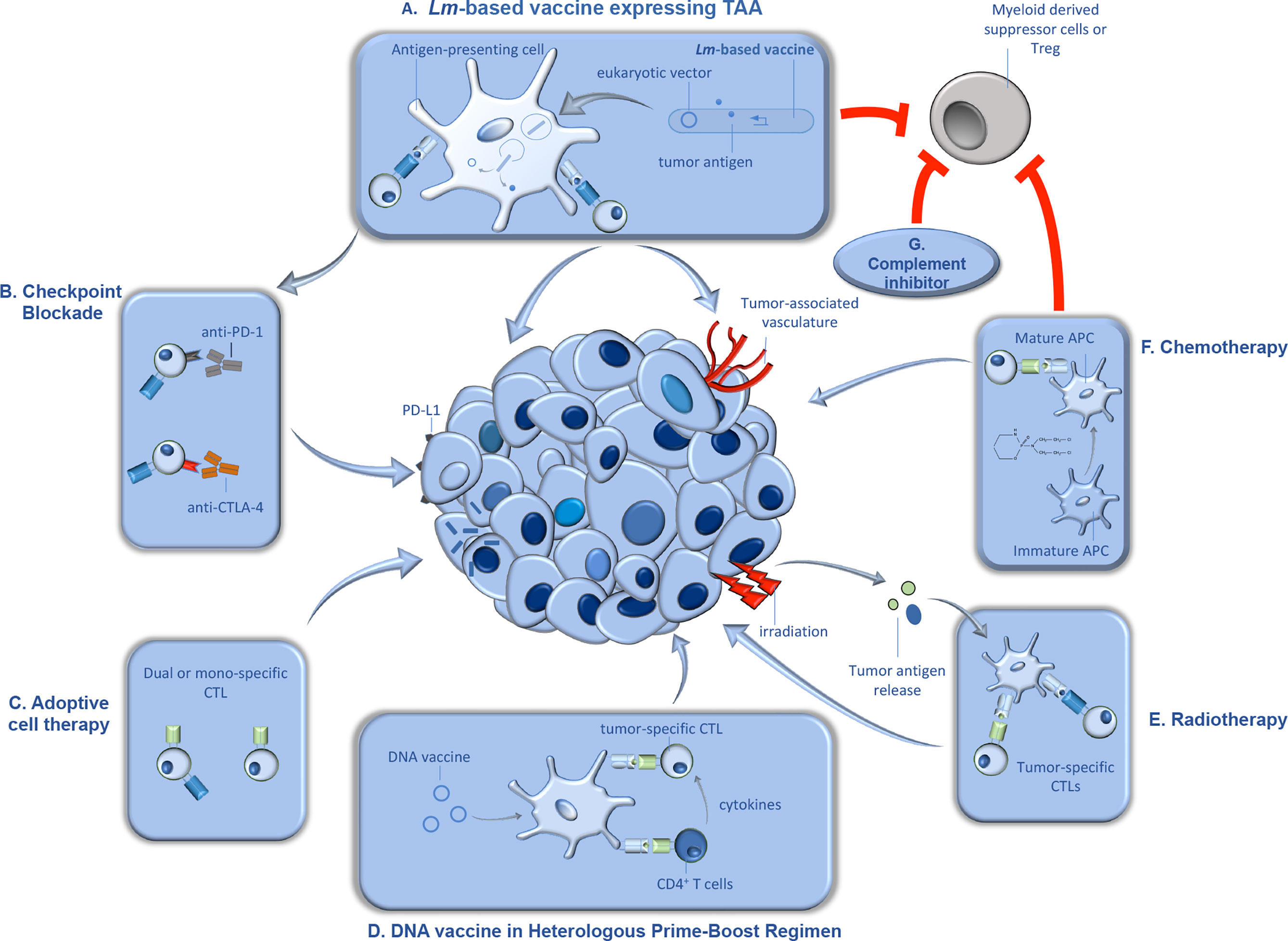
| 01:55, 15 April 2024 | Lmvaccine.jpg (file) |  |
436 KB | Chu2 | 1 | |
| 01:54, 15 April 2024 | Screenshot 2024-04-14 at 9.53.54 PM.png (file) |  |
1.42 MB | Mccune2 | 1 | |
| 01:54, 15 April 2024 | Botox2.png (file) |  |
1.34 MB | Leclerc1 | 1 | |
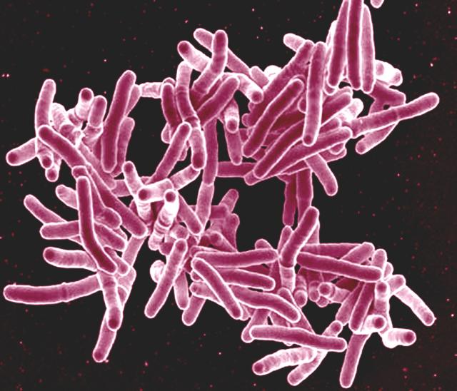
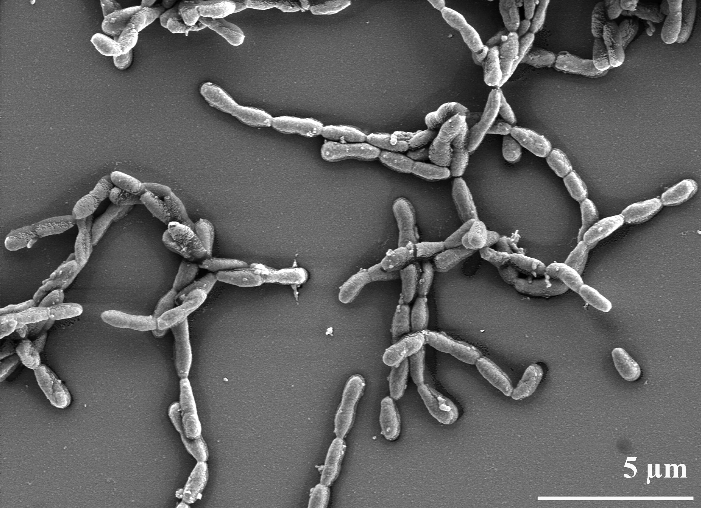
| 01:51, 15 April 2024 | M. tuberculosis.jpg (file) |  |
57 KB | Schwingel | Mycobacterium Tuberculosis Bacteria scanning electron micrograph Scanning electron micrograph of Mycobacterium tuberculosis bacteria. Credit: NIAID. Licensed under CC BY 2.0 DEED https://www.flickr.com/photos/niaid/5149398656 | 1 |
| 01:46, 15 April 2024 | Variant phenotyple table.jpg (file) |  |
60 KB | Chu2 | 1 | |
| 01:45, 15 April 2024 | Blackmap.png (file) |  |
304 KB | Breard1 | 1 | |
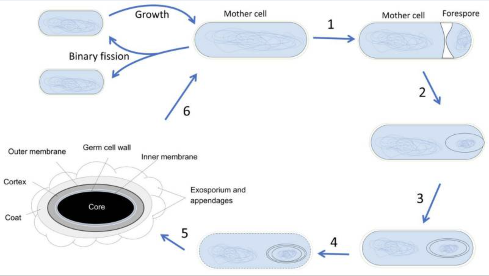
| 01:45, 15 April 2024 | Spore2.png (file) |  |
718 KB | Leclerc1 | 1 | |
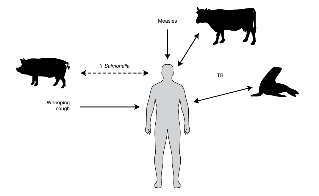
| 01:45, 15 April 2024 | Stone Domestic Origins Hypothesis.png (file) |  |
60 KB | Schwingel | The domestic origins for human disease hypothesis. This posits that agriculturalists living in close proximity with animals, especially livestock, would have been at high risk for zoonotic pathogens. From Stone, Anne C. 2020. “Getting Sick in the Neolithic.” Nature Ecology & Evolution 4 (3): 286–87. https://doi.org/10.1038/s41559-020-1115-8. | 1 |
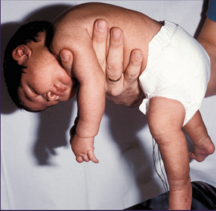
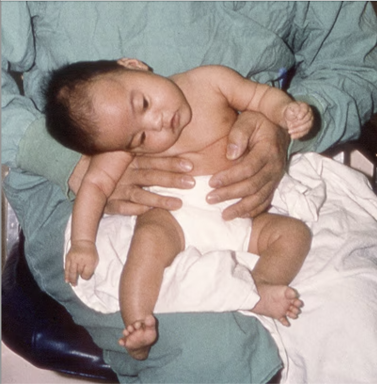
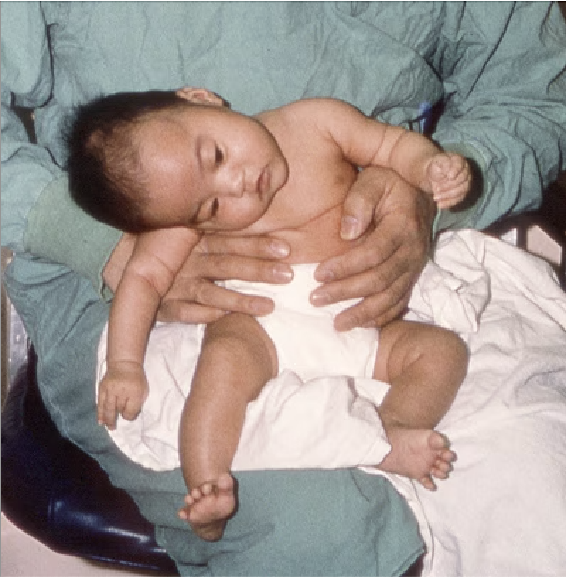
| 01:44, 15 April 2024 | Baby2.png (file) |  |
391 KB | Leclerc1 | 1 | |
| 01:42, 15 April 2024 | Dyado.jpg (file) |  |
536 KB | Hconawa | 1 | |
| 01:42, 15 April 2024 | White24.png (file) |  |
61 KB | Breard1 | 1 | |
| 01:39, 15 April 2024 | Gray.jpg (file) |  |
205 KB | Leclerc1 | 1 | |
| 01:39, 15 April 2024 | Map5.png (file) |  |
61 KB | Breard1 | 1 | |
| 01:36, 15 April 2024 | Kid.png (file) |  |
390 KB | Leclerc1 | 1 | |
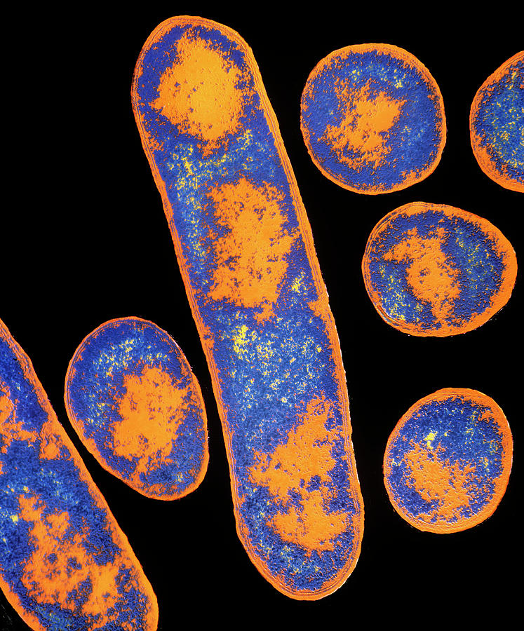
| 01:35, 15 April 2024 | Https---media.sciencephoto.com-image-b2200365-800wm-B2200365-False-colour TEM of Clostridium botulinum.jpg (file) |  |
296 KB | Leclerc1 | 1 | |
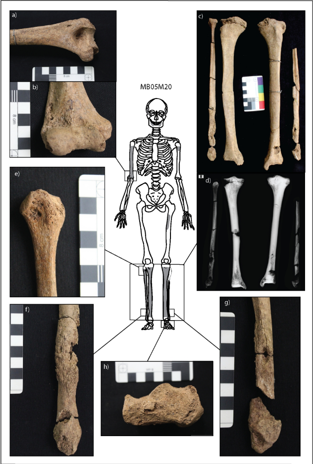
| 01:33, 15 April 2024 | Vlok et al. Fig 2 M20.png (file) |  |
788 KB | Schwingel | Lesions related to infectious disease in individual M20 from Neolithic Northern Vietnam. A) and b) show cavitations (holes) in the right humerus. C) and d) show new bone on the tibiae and fibulae. E), f), and g) show new bone growth and cavitations on the right fibula. H) shows cavitations on the left heel bone. From Vlok, Melandri, Marc Oxenham, Kate Domett, Tran Thi Minh, Thi Mai Huong Nguyen, Hirofumi Matsumura, Hiep Hoang Trinh, et al. 2020. “Two Probable Cases of Infection with Treponema... | 1 |
| 01:31, 15 April 2024 | EBOVpic.png (file) |  |
61 KB | Breard1 | 1 | |
| 01:31, 15 April 2024 | Microbial interactions.webp (file) |  |
112 KB | Epperson1 | 1 | |
| 01:22, 15 April 2024 | Bats1234.jpg (file) |  |
15 KB | Breard1 | 1 | |
| 01:15, 15 April 2024 | Bat123.jpeg (file) |  |
24 KB | Breard1 | 1 | |
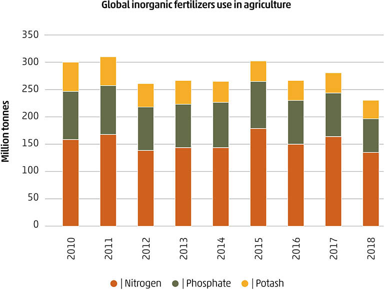
| 01:13, 15 April 2024 | Fertilizer graph.jpeg (file) |  |
65 KB | Epperson1 | 1 | |
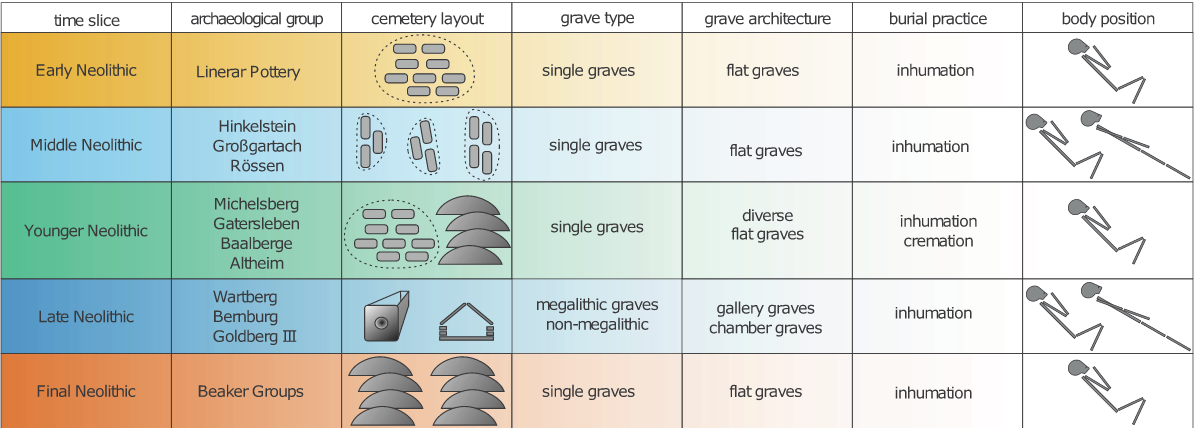
| 01:13, 15 April 2024 | Fuchs et al. Mortuary Practices Neolithic.png (file) |  |
214 KB | Schwingel | Fuchs et al. Figure 3: Archaeological groups, burials, and mortuary practices throughout the Neolithic in Germany. Image from Fuchs, Katharina, Christoph Rinne, Clara Drummer, Alexander Immel, Ben Krause-Kyora, and Almut Nebel. “Infectious Diseases and Neolithic Transformations: Evaluating Biological and Archaeological Proxies in the German Loess Zone between 5500 and 2500 BCE.” 2019. The Holocene 29 (10): 1545–57.http://journals.sagepub.com/doi/10.1177/0959683619857230 | 1 |
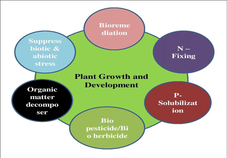
| 01:08, 15 April 2024 | Plant growth.png (file) |  |
108 KB | Epperson1 | 1 | |
| 01:02, 15 April 2024 | Low.png (file) |  |
144 KB | Johnson14 | 1 | |
| 01:02, 15 April 2024 | Din.png (file) |  |
433 KB | Johnson14 | 1 | |
| 01:00, 15 April 2024 | Din2.png (file) |  |
551 KB | Johnson14 | 1 |
