Clostridium as a Cancer Therapy
Clostridium is one of the largest prokaryotic genera and consists of a diverse range of obligatory anaerobic bacteria. Most of these bacteria are flagellated and motile. These bacteria are Gram-positive and rod-shaped, and capable of forming endospores. Clostridium bacteria can undergo a complex cell differentiation process that produces endospores that are highly resistant to harsh environmental conditions and can withstand high temperature, disinfectants, and low-energy radiation. The genus includes common free-living bacteria and pathogens. Several pathogens producing potent toxins are part of this genera including C. tetani, C. botulinum, C. difficile, and C. perfringens. C. tetani and C. botulinum are both well-studied species linked to human diseases and are potent bacterial toxins. C. tetani is the causative agent of tetanus, and Clostridium botulinum is related to food poisoning and causes botulism. Other members of the Clostridium genus include Clostridium perfringens which can infect wounds and cause gas gangrene, and Clostridium difficile, which grows in the gut during antibiotic therapy to cause pseudomembranous enterocolitis. Recently, Clostridium novyi type A has been associated with an outbreak of serious illness and death amongst intravenous drug users. However, most Clostridium species are nonpathogenic. Many are harmless and can be found in the soil. Some species have applications in bioremediation and wastewater treatment. Nonpathogenic species include the industrially valuable Clostridium acetobutylicum and Clostridium beijerinckii. These solventogenic clostridia are used in acetone, butanol and isopropanol fermentation.
Section 1
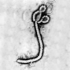
At right is a sample image insertion. It works for any image uploaded anywhere to MicrobeWiki. The insertion code consists of:
Double brackets: [[
Filename: Ebola virus 1.jpeg
Thumbnail status: |thumb|
Pixel size: |300px|
Placement on page: |right|
Legend/credit: Electron micrograph of the Ebola Zaire virus. This was the first photo ever taken of the virus, on 10/13/1976. By Dr. F.A. Murphy, now at U.C. Davis, then at the CDC.
Closed double brackets: ]]
Other examples:
Bold
Italic
Subscript: H2O
Superscript: Fe3+
Overall paper length should be 3,000 words, with at least 3 figures with data.
Section 2
Include some current research in each topic, with at least one figure showing data.
Section 3
Include some current research in each topic, with at least one figure showing data.
Further Reading
[Sample link] Ebola Hemorrhagic Fever—Centers for Disease Control and Prevention, Special Pathogens Branch
References
Edited by Anh Tran, a student of Nora Sullivan in BIOL168L (Microbiology) in The Keck Science Department of the Claremont Colleges Spring 2014.

