Bacteroides fragilis: Difference between revisions
(Page created) |
No edit summary |
||
| Line 2: | Line 2: | ||
==Introduction== | ==Introduction== | ||
<i>Bacteroides fragilis</i> is a Gram-negative bacterium found in the human colon (1). It is responsible for a large number of opportunistic infections in hospitals and contributes significantly to morbidity and mortality (2). <i>B. fragilis</i> is of interest to researchers because of its ability to evade immune responses and evolving drug resistance. | |||
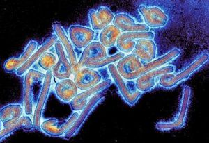
[[Image:marburgvirus.jpg|thumb|300px|right|Colony of Marburg virus. Transmission electron microscope image taken by Dr. Tom Geisbert]] | [[Image:marburgvirus.jpg|thumb|300px|right|Colony of Marburg virus. Transmission electron microscope image taken by Dr. Tom Geisbert]] | ||
Revision as of 11:29, 8 November 2019
Introduction
Bacteroides fragilis is a Gram-negative bacterium found in the human colon (1). It is responsible for a large number of opportunistic infections in hospitals and contributes significantly to morbidity and mortality (2). B. fragilis is of interest to researchers because of its ability to evade immune responses and evolving drug resistance.
At right is a sample image insertion. It works for any image uploaded anywhere to MicrobeWiki. The insertion code consists of:
Double brackets: [[
Filename: PHIL_1181_lores.jpg
Thumbnail status: |thumb|
Pixel size: |300px|
Placement on page: |right|
Legend/credit: Electron micrograph of the Ebola Zaire virus. This was the first photo ever taken of the virus, on 10/13/1976. By Dr. F.A. Murphy, now at U.C. Davis, then at the CDC.
Closed double brackets: ]]
Other examples:
Bold
Italic
Subscript: H2O
Superscript: Fe3+
Section 1 Genetics
Include some current research, with at least one image.
Sample citations: [1]
[2]
A citation code consists of a hyperlinked reference within "ref" begin and end codes.
Section 2 Microbiome
Include some current research, with a second image.
Conclusion
Overall text length should be at least 1,000 words (before counting references), with at least 2 images. Include at least 5 references under Reference section.
References
Edited by James Cawthon, student of Joan Slonczewski for BIOL 116 Information in Living Systems, 2019, Kenyon College.

