File:Infected Daphnia healthy Daphnia and SEM Metschnikowia spores.jpeg: Difference between revisions
From MicrobeWiki, the student-edited microbiology resource
m (Kgriebel moved page File:Infected Daphnia healthy Daphnia and TEM Metschnikowia spores.jpeg to File:Infected Daphnia healthy Daphnia and SEM Metschnikowia spores.jpeg: scanning electron (SEM) not tunneling electron (TEM)) |
No edit summary |
||
| Line 1: | Line 1: | ||
From left, an infected Daphnia dentifera, an uninfected D. dentifera, and an infected D. dentifera. Photo by A. J. Tessier. On right, a scanning electron microscopy image showing M. bicuspidata spores as they appear under the ruptured carapace of an infected zooplankter. Photo by Carol Flegler. (Hall et al., 2006). | |||
Latest revision as of 23:11, 27 April 2015
From left, an infected Daphnia dentifera, an uninfected D. dentifera, and an infected D. dentifera. Photo by A. J. Tessier. On right, a scanning electron microscopy image showing M. bicuspidata spores as they appear under the ruptured carapace of an infected zooplankter. Photo by Carol Flegler. (Hall et al., 2006).
File history
Click on a date/time to view the file as it appeared at that time.
| Date/Time | Thumbnail | Dimensions | User | Comment | |
|---|---|---|---|---|---|
| current | 23:06, 27 April 2015 | 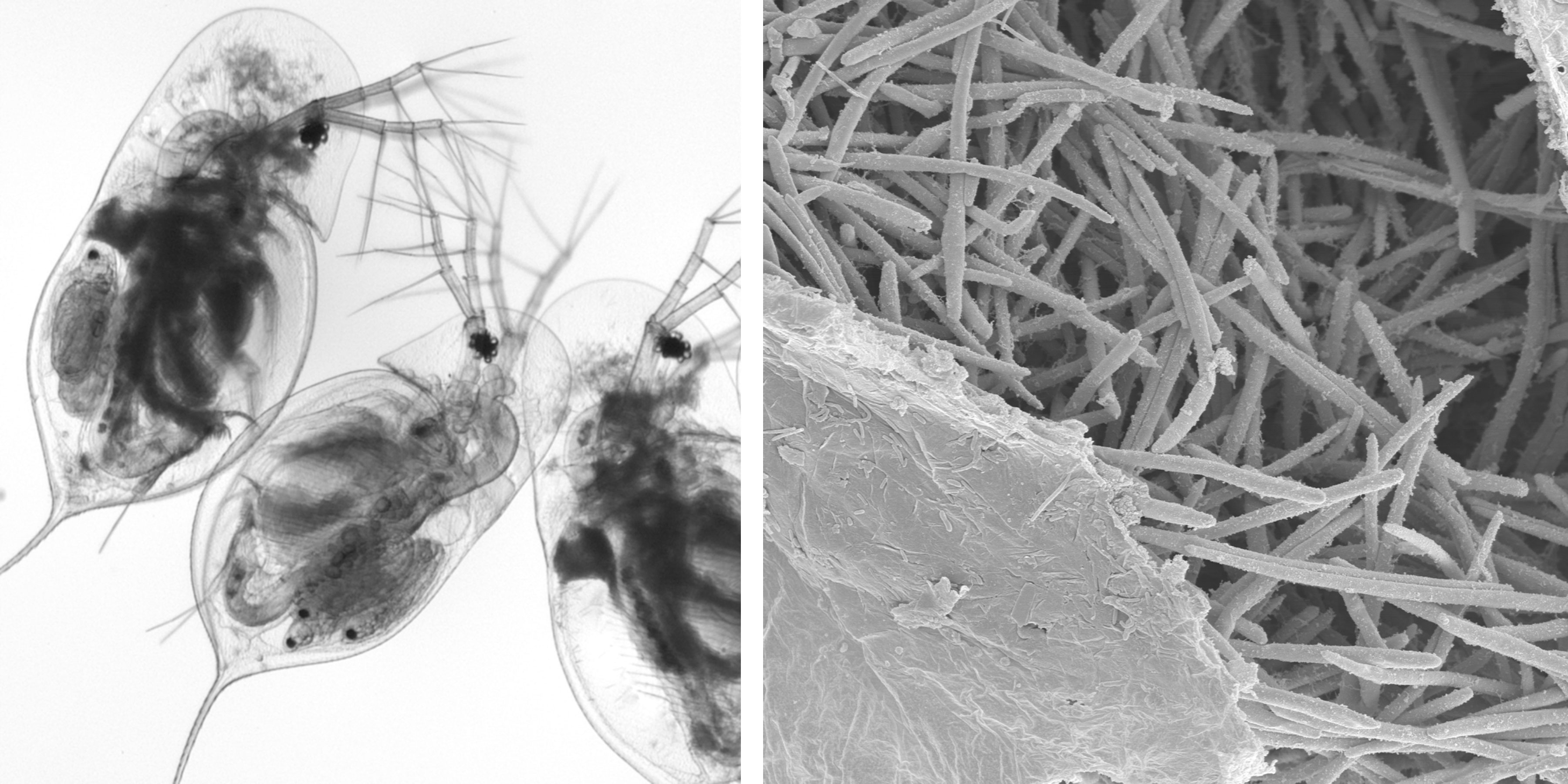 | 3,600 × 1,800 (733 KB) | Kgriebel (talk | contribs) | From left, an infected Daphnia, an uninfected Daphnia, and an infected Daphnia. Photo by A. J. Tessier. On right, a scanning electron microscopy image showing spores as they appear under the ruptured carapace of a Daphnia. Photo by Carol Flegler. (H... |
| 19:37, 26 April 2015 |  | 3,600 × 1,800 (733 KB) | Kgriebel (talk | contribs) | From left, an infected Daphnia, an uninfected Daphnia, and an infected Daphnia. On right, an electron microscopy image showing spores as they appear under the ruptured carapace of a Daphnia. |
You cannot overwrite this file.
File usage
The following 3 pages use this file:
