File:BaylisascarisCerebralSection.png
From MicrobeWiki, the student-edited microbiology resource

Size of this preview: 800 × 541 pixels. Other resolution: 1,838 × 1,244 pixels.
Original file (1,838 × 1,244 pixels, file size: 4.09 MB, MIME type: image/png)
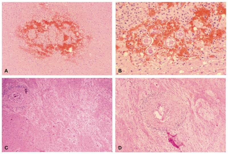
This image from Kazacos et al. (2013) shows the migration track in the cerebrum of a monkey infected by B. procyonis (A, B), as well as in a child (C,D). In both cases, infection was fatal. Invading macrophages can be seen in ,A, and a larva can be seen in B. In C, a larval granuloma is visible, and is magnified more closely in D.” From http://www.sciencedirect.com/science/article/pii/B9780444534903000200.
File history
Click on a date/time to view the file as it appeared at that time.
| Date/Time | Thumbnail | Dimensions | User | Comment | |
|---|---|---|---|---|---|
| current | 01:28, 20 April 2017 | | 1,838 × 1,244 (4.09 MB) | Torokn (talk | contribs) | This image from Kazacos et al. (2013) shows the migration track in the cerebrum of a monkey infected by <i>B. procyonis</i> (A, B), as well as in a child (C,D). In both cases, infection was fatal. Invading macrophages can be seen in ,<i>A</i>, and a la... |
You cannot overwrite this file.
File usage
The following page uses this file:
