File:Figure 1.PNG
From MicrobeWiki, the student-edited microbiology resource

Size of this preview: 464 × 600 pixels. Other resolution: 935 × 1,209 pixels.
Original file (935 × 1,209 pixels, file size: 347 KB, MIME type: image/png)
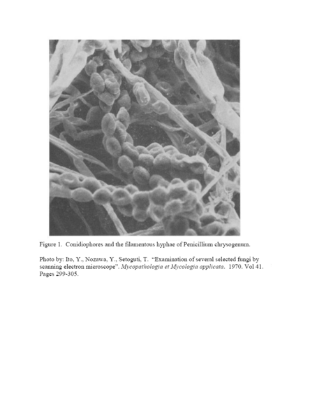
Figure 1. Conidiophores and the filamentous hyphae of Penicillium chrysogenum.
Photo by: Ito, Y., Nozawa, Y., Setoguti, T. “Examination of several selected fungi by scanning electron microscope”. Mycopathologia et Mycologia applicata. 1970. Vol 41. Pages 299-305.
File history
Click on a date/time to view the file as it appeared at that time.
| Date/Time | Thumbnail | Dimensions | User | Comment | |
|---|---|---|---|---|---|
| current | 00:58, 27 August 2007 | | 935 × 1,209 (347 KB) | Goattfd (talk | contribs) | |
| 00:55, 27 August 2007 |  | 935 × 1,209 (335 KB) | Goattfd (talk | contribs) | Reverted to earlier revision | |
| 00:54, 27 August 2007 |  | 935 × 1,209 (372 KB) | Goattfd (talk | contribs) | Reverted to earlier revision | |
| 23:31, 26 August 2007 |  | 935 × 1,209 (335 KB) | Goattfd (talk | contribs) | Figure 1. Conidiophores and the filamentous hyphae of Penicillium chrysogenum. Photo by: Ito, Y., Nozawa, Y., Setoguti, T. “Examination of several selected fungi by scanning electron microscope”. Mycopathologia et Mycologia applicata. 1970. Vol | |
| 23:29, 26 August 2007 |  | 935 × 1,209 (372 KB) | Goattfd (talk | contribs) | ||
| 23:27, 26 August 2007 |  | 935 × 1,209 (372 KB) | Goattfd (talk | contribs) | ||
| 23:26, 26 August 2007 |  | 935 × 1,209 (372 KB) | Goattfd (talk | contribs) |
You cannot overwrite this file.
File usage
The following page uses this file:
