File:JuninVirus.png

Original file (1,728 × 2,304 pixels, file size: 252 KB, MIME type: image/png)
Summary
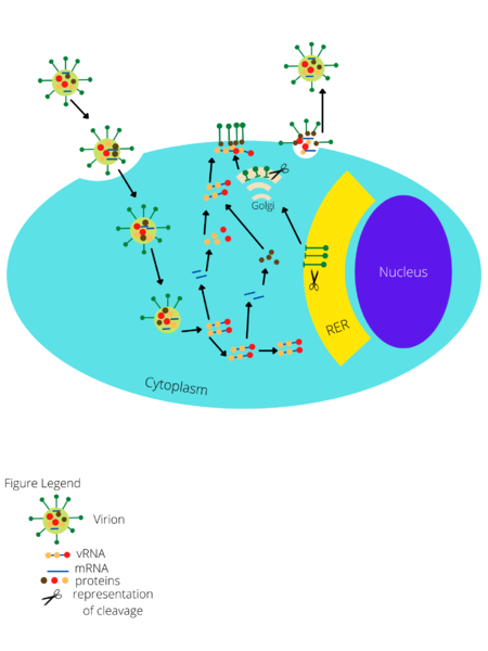
Figure 1: Visual representation of Junin Virus genome structure and life cycle. Adapted from Review of Mammarenavirus Biology and Replication[8] The virion first enters the host cell through endocytosis and then the RNA inside of it is released. There are two stages of mRNA: early and late. Early mRNA goes through NP and LP translation while late mRNA goes through Z translation. After translation, the proteins join together and the virion is assembled and released from the cell. Alongside this genomic viral RNA is synthesized and in the rough endoplasmic reticulum and Golgi body glycosylation and cleavage of GP1/GP2 occurs.
File history
Click on a date/time to view the file as it appeared at that time.
| Date/Time | Thumbnail | Dimensions | User | Comment | |
|---|---|---|---|---|---|
| current | 14:46, 6 December 2021 |  | 1,728 × 2,304 (252 KB) | Shahs07 (talk | contribs) | Figure 1: Visual representation of Junin Virus genome structure and life cycle. Adapted from Review of Mammarenavirus Biology and Replication[8] The virion first enters the host cell through endocytosis and then the RNA inside of it is released. There are two stages of mRNA: early and late. Early mRNA goes through NP and LP translation while late mRNA goes through Z translation. After translation, the proteins join together and the virion is assembled and released from the cell. Alongside thi... |
You cannot overwrite this file.
File usage
The following page uses this file:
