File:New Sulfur Bacteria Microscopy 1.png
From MicrobeWiki, the student-edited microbiology resource
No higher resolution available.
New_Sulfur_Bacteria_Microscopy_1.png (237 × 440 pixels, file size: 88 KB, MIME type: image/png)
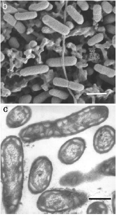
Scanning Electron Microscopy of filter-deposited cells (B) and thin section Transmission Electron Microscopy (C). Note the electron transparent structures near the perimeter of the cell. These structures are characteristic of of chlorosomes. Bar=300nm.Image courtesy of Beatty, Blankenship, et al. (2005).
File history
Click on a date/time to view the file as it appeared at that time.
| Date/Time | Thumbnail | Dimensions | User | Comment | |
|---|---|---|---|---|---|
| current | 04:06, 16 April 2009 | | 237 × 440 (88 KB) | Eldahank (talk | contribs) | Scanning Electron Microscopy of filter-deposited cells (B) and thin section Transmission Electron Microscopy (C). Note the electron transparent structures near the perimeter of the cell. These structures are characteristic of of chlorosomes. Bar=300nm.Ima |
You cannot overwrite this file.
File usage
The following page uses this file:
