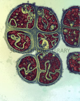
Micrococcus tetragenus
Classification
Kingdom - Bacteria; Phylum - Actinobacteria; Class - Actinobacteria; Order - Actinomycetales; Family - Micrococcaceae
Micrococcus tetragenus
Description and Significance
M. tetragenus is part of the Micrococcacae family. It is Gram positive and retains a spherical shape. It may be found in single or irregular groups of cells. [2] It's size varies from 0.5 to 3 micrometers in diameter. [2]This microbe is important because of its unique septation which is discussed in the following section. [3] M. tetragenus has also been shown to cause septicemia. [4] This microbe is similar to others in its group in shape and environment, although it has the unique septum and known pathogenesis. [4]
Structure, Metabolism, and Life Cycle
By far the most interesting feature about M. tetragenus is related to both its structure and life cylce. During septation, a cell wall and envelope form a septum that will be part of the new daughter cells. [3]. Regarding spherical cells, if the septum forms a multiple angles, the closely related staphylococci will result. In the case of Micrococcus tetragenus, a tetrad is formed with two septum are formed at right angles. [3]. The photograph included in this page shows the tetrad being formed. [2]. Micrococcus are known for their high G-C count and part of their metabolism allows them to produce acid from glucose glycerol and convert nitrate to nitrite. They are motile bacteria. [5]
Ecology and Pathogenesis
The natural habitat of Micrococcus tetragenus is in humans, usually found in the nasopharynx. It was also found in tuberculous cavities of the lungs in patients with TB. [4]
M. tetragenus is a cause of pulmonary infections. [3]. It has been documented to cause septicemia, with varying symptoms, most commonly upper respiratory infections. [4]. Interesting, during WWI, there were noted to be two small epidemics among soldiers where at least 25 had septicemia. [4]. This microbe is not usually the first cause of infection and therefore is known as a secondary invader. [4]
References
[1] NCBI Taxonomy. US Government, 28 Oct. 2009. Web. 22 July 2013. <http://www.ncbi.nlm.nih.gov/Taxonomy/Browser/wwwtax.cgi?mode=Info&id=1269&lvl=3&lin=f&keep=1&srchmode=1&unlock>.
[2] KWANGSHIN KIM/SCIENCE PHOTO LIBRARY, . Micrococcus tetragenus. 2013. Science Photo Library Ltd., London, UK. Web. 21 July 2013. <http://www.sciencephoto.com/media/12343/view>.
[3] Slonczewski, Joan L., and John W. Foster. Microbiology: An Evolving Science. 2ndnd ed. N.p.: n.p., 2009. 103-05. Print.
[4] Reimann, Hobart A. "Micrococcus tetragenus Infection." Department of Medicine, University of Minnesota Hospital, Minneapolis (1934): 311-19. Web. 22 July 2013. <http://www.jci.org/articles/view/100679/files/pdf>.
[5] Smith, K. J., R. Neafie, J. Yeager, and H. G. Skelton. 1999. "Micrococcus folliculitis in HIV-1 disease." British Journal of Dermatology, vol. 141, no. 3. British Association of Dermatologists. (558-561)
Author
Page authored by Samuel Goldenberg, student of Mandy Brosnahan, Instructor at the University of Minnesota-Twin Cities, MICB 3301/3303: Biology of Microorganisms.

