Chronic Bee Paralysis Virus: Difference between revisions
No edit summary |
No edit summary |
||
| (9 intermediate revisions by 2 users not shown) | |||
| Line 1: | Line 1: | ||
{{ | {{Moore2013}} | ||
{{Viral Biorealm Family}} | {{Viral Biorealm Family}} | ||
<br><br><br> | <br><br><br> | ||
| Line 6: | Line 6: | ||
==Baltimore Classification== | ==Baltimore Classification== | ||
Group IV: (+) ssRNA virus <sup>[[#References|[1]]]</sup> | [http://en.wikipedia.org/wiki/Baltimore_classification#Class_IV_.26_V:_Single-stranded_RNA_viruses Group IV:] (+) ssRNA virus <sup>[[#References|[1]]]</sup> | ||
==Higher order categories== | ==Higher order categories== | ||
| Line 24: | Line 24: | ||
The complete sequences of the two RNAs of Chronic bee paralysis virus (CBPV) have been determined: RNA 1 (3674nt long) and RNA 2 (2305nt long) are positive single-stranded RNAs that are capped but not polyadenylated <sup>[[#References|[1]]] </sup>. Figure 2 shows the two RNAs discovered and their orientation <sup>[[#References|[5]]]</sup>. | The complete sequences of the two RNAs of Chronic bee paralysis virus (CBPV) have been determined: RNA 1 (3674nt long) and RNA 2 (2305nt long) are positive single-stranded RNAs that are capped but not polyadenylated <sup>[[#References|[1]]] </sup>. Figure 2 shows the two RNAs discovered and their orientation <sup>[[#References|[5]]]</sup>. | ||
Initial research showed the presence of two major and 3 minor RNAs. However, further studies showed that the 3 minor RNAs may have been what is now believed to be the Chronic bee paralysis virus satellite <sup>[[#References|[5]]]</sup>. | Initial research showed the presence of two major and 3 minor RNAs. However, further studies showed that the 3 minor RNAs may have been what is now believed to be the Chronic bee paralysis virus satellite <sup>[[#References|[5]]]</sup>. | ||
[[File:Phylo_treeB.jpeg|200px|thumb|right|Figure 3. | [[File:Phylo_treeB.jpeg|200px|thumb|right|Figure 3. Proposed phylogenetic tree showing the relationship between CPBV and two known families <sup>[[#References|[5]]] </sup>.]] | ||
The amino acid information obtained from the ORF 3 on RNA 1 shared the conserved motifs of the RNA-dependent RNA polymerase (RdRp) sequence and presented similarities with other known families. However, no similarities were found between the other CBPV amino acid sequences and sequences in the NCBI databases, suggesting that CBPV is a new family of positive single-stranded RNA viruses <sup>[[#References|[1]]]</sup>. Based on this similarity, the phylogenetic position of CBPV has been placed somewhere between ''Nodaviridae'' and ''Tombusviridae'' (see Fig. 3). | The amino acid information obtained from the ORF 3 on RNA 1 shared the conserved motifs of the RNA-dependent RNA polymerase (RdRp) sequence and presented similarities with other known families. However, no similarities were found between the other CBPV amino acid sequences and sequences in the NCBI databases, suggesting that CBPV is a new family of positive single-stranded RNA viruses <sup>[[#References|[1]]]</sup>. Based on this similarity, the phylogenetic position of CBPV has been placed somewhere between ''Nodaviridae'' and ''Tombusviridae'' (see Fig. 3). | ||
| Line 30: | Line 30: | ||
[[File:CellB.jpeg|100px|thumb|left|Figure 4. Electron microscopy image showing CBPV particles purified from experimentally infected bees <sup> [[#References|[5]]]</sup>.]] | [[File:CellB.jpeg|100px|thumb|left|Figure 4. Electron microscopy image showing CBPV particles purified from experimentally infected bees <sup> [[#References|[5]]]</sup>.]] | ||
CBPV is somewhat unusual. The particles have been found to vary in size and structure (see Fig. 4). Purified preparations of the virus contain non-enveloped, anisometric, ellipsoidal particles, 30-65 nm in length and 20 nm in width <sup>[[#References|[5]]]</sup>. However, other shapes have been observed as well: rings, figure eights, branching rods and lengths up to 640 nm. | The structure of CBPV is somewhat unusual. The particles have been found to vary in size and structure (see Fig. 4). Purified preparations of the virus contain non-enveloped, anisometric, ellipsoidal particles, 30-65 nm in length and 20 nm in width <sup>[[#References|[5]]]</sup>. However, other shapes have been observed as well: rings, figure eights, branching rods and lengths up to 640 nm. | ||
The virus particles contain a single capsid protein with a size of 23.5 kDa <sup>[[#References|[5]]]</sup>. Additional studies are required to further characterize the virus structure and its proteins. | The virus particles contain a single capsid protein with a size of 23.5 kDa <sup>[[#References|[5]]]</sup>. Additional studies are required to further characterize the virus structure and its proteins. | ||
| Line 46: | Line 46: | ||
''Type 1'' | ''Type 1'' | ||
The Type 1 syndrome is generally seen as the most common type <sup>[[#References|[5]]]</sup>. Bees infected with | The Type 1 syndrome is generally seen as the most common type <sup>[[#References|[5]]]</sup>. Bees infected with Type 1 are unable to fly and are seen crawling on the ground or on plants, sometimes in large groups of thousands of individuals. Other symptoms include bloated abdomen, caused by distension of the honey sac with fluid, and partially-open dislocated wings <sup>[[#References|[5]]]</sup>. Within a few days of infection, the bees die which can cause a huge detriment to the productivity of the hive. These symptoms are identical to symptoms attributed to the “Isle of Wight disease” seen in Britain in the early 1900s <sup>[[#References|[5]]]</sup>. | ||
''Type 2'' | ''Type 2'' | ||
| Line 52: | Line 52: | ||
At first, bees affected with the Type 2 syndrome can still fly, but they become hairless which causes them to appear darker, shinier and smaller than healthy bees (see Fig. 1 and Fig 5A) <sup>[[#References|[5]]]</sup>. As a result, healthy bees attack the affected – nibbling them as if they were robber bees. Within a few days of infection, the bees become flightless, begin to tremble and ultimately die <sup>[[#References|[5]]]</sup>. | At first, bees affected with the Type 2 syndrome can still fly, but they become hairless which causes them to appear darker, shinier and smaller than healthy bees (see Fig. 1 and Fig 5A) <sup>[[#References|[5]]]</sup>. As a result, healthy bees attack the affected – nibbling them as if they were robber bees. Within a few days of infection, the bees become flightless, begin to tremble and ultimately die <sup>[[#References|[5]]]</sup>. | ||
Given that other bee viruses also cause some of these same symptoms, diagnosis cannot rely on visual inspection of symptoms alone <sup>[[#References|[5]]]</sup>. However, it has also been discovered that seemingly healthy bees were infected and carried CBPV <sup>[[#References|[3]]]</sup>. | Given that other bee viruses also cause some of these same symptoms, diagnosis cannot rely on visual inspection of symptoms alone <sup>[[#References|[5]]]</sup>. However, it has also been discovered that seemingly healthy bees were infected and carried CBPV <sup>[[#References|[3]]]</sup>. The reason the virus can sometimes go unnoticed in a host is still unknown. | ||
==Future Studies & Global Impacts== | ==Future Studies & Global Impacts== | ||
Future research will attempt to better understand the method of infection and the reproduction of the virus within the host<sup>[[#References|[5]]]</sup>. Information that will help get to that end includes information on viral proteins and structure, worldwide infection rates and sequencing data. If this virus and other bee viruses continue to ravage bees, the impact could be unrecoverable detriments in bee populations. | Future research will attempt to better understand the method of infection and the reproduction of the virus within the host <sup>[[#References|[5]]]</sup>. Information that will help get to that end includes information on viral proteins and structure, worldwide infection rates and sequencing data. If this virus and other bee viruses continue to ravage bees, the impact could be unrecoverable detriments in bee populations. | ||
==References== | ==References== | ||
[1] Olivier, V.; Blanchard, P.; Chaouch, S.; Lallemand, P.; Schurr, F.; Celle, O.; Dubois, E.; Tordo, N.; Thiéry, R.; Houlgatte, R.; Ribière, M. Molecular characterisation and phylogenetic analysis of Chronic bee paralysis virus, a honeybee virus. Virus Research. 2008 March, Vol. 132 Issue 1/2, p59-68. DOI: 10.1016/j.virusres.2007.10.014. http://dx.doi.org/10.1016/j.virusres.2007.10.014 | * [1] Olivier, V.; Blanchard, P.; Chaouch, S.; Lallemand, P.; Schurr, F.; Celle, O.; Dubois, E.; Tordo, N.; Thiéry, R.; Houlgatte, R.; Ribière, M. Molecular characterisation and phylogenetic analysis of Chronic bee paralysis virus, a honeybee virus. Virus Research. 2008 March, Vol. 132 Issue 1/2, p59-68. DOI: 10.1016/j.virusres.2007.10.014. http://dx.doi.org/10.1016/j.virusres.2007.10.014 | ||
[2] Celle, O; Blanchard, P,; Olivier, V.; Schurr, F.; Cougoule, N.; Faucon, J-P.; Ribière, M. Detection of Chronic bee paralysis virus (CBPV) genome and its replicative RNA form in various hosts and possible ways of spread, Virus Research, Volume 133, Issue 2, May 2008, Pages 280-284, ISSN 0168-1702. DOI: 10.1016/j.virusres.2007.12.011. http://dx.doi.org/10.1016/j.virusres.2007.12.011 | * [2] Celle, O; Blanchard, P,; Olivier, V.; Schurr, F.; Cougoule, N.; Faucon, J-P.; Ribière, M. Detection of Chronic bee paralysis virus (CBPV) genome and its replicative RNA form in various hosts and possible ways of spread, Virus Research, Volume 133, Issue 2, May 2008, Pages 280-284, ISSN 0168-1702. DOI: 10.1016/j.virusres.2007.12.011. http://dx.doi.org/10.1016/j.virusres.2007.12.011 | ||
[3] Morimoto, T.; Kojima, Y.; Yoshiyama, M.; Kimura, K.; Yang, B.; Kadowaki, T. Molecular Identification of Chronic Bee Paralysis Virus Infection in Apis mellifera Colonies in Japan. Viruses. July, 2012. Vol. 4 Issue 7, p1093-1103. DOI: 10.3390/v4071093. http://dx.crossref.org/10.3390%2Fv4071093 | * [3] Morimoto, T.; Kojima, Y.; Yoshiyama, M.; Kimura, K.; Yang, B.; Kadowaki, T. Molecular Identification of Chronic Bee Paralysis Virus Infection in Apis mellifera Colonies in Japan. Viruses. July, 2012. Vol. 4 Issue 7, p1093-1103. DOI: 10.3390/v4071093. http://dx.crossref.org/10.3390%2Fv4071093 | ||
[4] Ribière, M. M., Lallemand, P. P., Iscache, A. L., Schurr, F. F., Celle, O., Blanchard, P. P., Olivier, V; Faucon, J. P. (2007). Spread of Infectious Chronic Bee Paralysis Virus by Honeybee (Apis mellifera L.) Feces. Applied & Environmental Microbiology, 73(23), 7711-7716. doi:10.1128/AEM.01053-07 | * [4] Ribière, M. M., Lallemand, P. P., Iscache, A. L., Schurr, F. F., Celle, O., Blanchard, P. P., Olivier, V; Faucon, J. P. (2007). Spread of Infectious Chronic Bee Paralysis Virus by Honeybee (Apis mellifera L.) Feces. Applied & Environmental Microbiology, 73(23), 7711-7716. doi:10.1128/AEM.01053-07 | ||
[5] Ribière, M.; Olivier, V.; Blanchard, P. Chronic bee paralysis: A disease and a virus like no other? Journal of Invertebrate Pathology, Volume 103, Supplement, January 2010, Pages S120–S131. http://dx.doi.org/10.1016/j.jip.2009.06.013 | * [5] Ribière, M.; Olivier, V.; Blanchard, P. Chronic bee paralysis: A disease and a virus like no other? Journal of Invertebrate Pathology, Volume 103, Supplement, January 2010, Pages S120–S131. http://dx.doi.org/10.1016/j.jip.2009.06.013 | ||
Edited by Naomi Chouinard of Dr. Lisa R. Moore, University of Southern Maine, Department of Biological Sciences, http://www.usm.maine.edu/bio | Edited by Naomi Chouinard of Dr. Lisa R. Moore, University of Southern Maine, Department of Biological Sciences, http://www.usm.maine.edu/bio | ||
Latest revision as of 15:17, 22 February 2016
A Viral Biorealm page on the family Chronic Bee Paralysis Virus
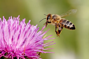
Chronic bee paralysis virus (often abbreviated CBPV or CPV) causes chronic bee paralysis disease in honeybees around the world [1]. This virus is one of many bee viruses that are becoming an increasing problem in the bee community. Chronic bee paralysis virus (CBPV) is the causal agent of chronic paralysis known to induce significant losses in honeybee colonies [2].
Baltimore Classification
Higher order categories
Virus; Positive single-stranded RNA virus. Although CBPV shares several characteristics with the Nodaviridae and Tombusviridae virus families, CBPV is considered a new family of positive single-stranded RNA viruses [3]. The virus is not fully classified.
Description and Significance
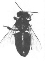
The common host for the chronic bee paralysis virus is the honeybee, of which Apis mellifera is the most common species. This virus has been found in various species around the world and is a problem because it causes large numbers of honeybee deaths [1]. Honeybees play and important role as pollinators to a large variety of plants, thus they hold an important role in agriculture. With higher rates of CBPV comes more bee deaths and lower plant production. This can have huge impacts on agriculture, both on the large and small scale. Keeping track of such viruses is important in foreign trade. In some cases, infected bees were passed through undetected to other countries during trade and infected colonies in that country with CBPV [3]. Proper detection and management techniques are key in preventing further spread of CBPV.
Symptoms of CBPV include severe trembling of the wings and body, crawling on the ground, loss of hair and a darker appearance (see Fig. 1 and Fig. 3) [3]. Most infected bees die within a few days of infection. However, it has also been seen that the virus can infect seemingly healthy bees. The virus is spread through direct bee to bee contact and perhaps contact with feces [4]. CBPV has also more recently been found in ants and mites [2], which could provide another method of spread: mite to bee contact.
Genome Structure
.
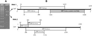
The complete sequences of the two RNAs of Chronic bee paralysis virus (CBPV) have been determined: RNA 1 (3674nt long) and RNA 2 (2305nt long) are positive single-stranded RNAs that are capped but not polyadenylated [1] . Figure 2 shows the two RNAs discovered and their orientation [5]. Initial research showed the presence of two major and 3 minor RNAs. However, further studies showed that the 3 minor RNAs may have been what is now believed to be the Chronic bee paralysis virus satellite [5].
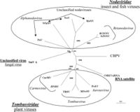
The amino acid information obtained from the ORF 3 on RNA 1 shared the conserved motifs of the RNA-dependent RNA polymerase (RdRp) sequence and presented similarities with other known families. However, no similarities were found between the other CBPV amino acid sequences and sequences in the NCBI databases, suggesting that CBPV is a new family of positive single-stranded RNA viruses [1]. Based on this similarity, the phylogenetic position of CBPV has been placed somewhere between Nodaviridae and Tombusviridae (see Fig. 3).
Virion Structure

The structure of CBPV is somewhat unusual. The particles have been found to vary in size and structure (see Fig. 4). Purified preparations of the virus contain non-enveloped, anisometric, ellipsoidal particles, 30-65 nm in length and 20 nm in width [5]. However, other shapes have been observed as well: rings, figure eights, branching rods and lengths up to 640 nm.
The virus particles contain a single capsid protein with a size of 23.5 kDa [5]. Additional studies are required to further characterize the virus structure and its proteins.
Reproductive Cycle in a Host Cell
The spread of the virus is done through contact with infected bees. Some studies show that the virus may be transmitted via bee feces [4]. Dissected regions of infected deceased bees were shown to contain viral loads of 1010-1012 copies, with the highest numbers in the head [5]. At the present level of research, little can be said about how the virus is established in the host, how it is activated, what form the virus stays in or the reactions between this virus and others [5].
Viral Ecology & Pathology
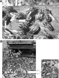
Studies have shown that CBPV remains viral when found in the feces of infected bees. CBPV was even passed on to healthy bees by being in the presence of infected bee feces alone [4]. Symptoms of CBPV include severe trembling of the wings and body, crawling on the ground, hairlessness, darker or shinier appearance, death, etc. CBPV causes chronic paralysis disease which presents itself as two syndromes with two sets of symptoms:
Type 1
The Type 1 syndrome is generally seen as the most common type [5]. Bees infected with Type 1 are unable to fly and are seen crawling on the ground or on plants, sometimes in large groups of thousands of individuals. Other symptoms include bloated abdomen, caused by distension of the honey sac with fluid, and partially-open dislocated wings [5]. Within a few days of infection, the bees die which can cause a huge detriment to the productivity of the hive. These symptoms are identical to symptoms attributed to the “Isle of Wight disease” seen in Britain in the early 1900s [5].
Type 2
At first, bees affected with the Type 2 syndrome can still fly, but they become hairless which causes them to appear darker, shinier and smaller than healthy bees (see Fig. 1 and Fig 5A) [5]. As a result, healthy bees attack the affected – nibbling them as if they were robber bees. Within a few days of infection, the bees become flightless, begin to tremble and ultimately die [5].
Given that other bee viruses also cause some of these same symptoms, diagnosis cannot rely on visual inspection of symptoms alone [5]. However, it has also been discovered that seemingly healthy bees were infected and carried CBPV [3]. The reason the virus can sometimes go unnoticed in a host is still unknown.
Future Studies & Global Impacts
Future research will attempt to better understand the method of infection and the reproduction of the virus within the host [5]. Information that will help get to that end includes information on viral proteins and structure, worldwide infection rates and sequencing data. If this virus and other bee viruses continue to ravage bees, the impact could be unrecoverable detriments in bee populations.
References
- [1] Olivier, V.; Blanchard, P.; Chaouch, S.; Lallemand, P.; Schurr, F.; Celle, O.; Dubois, E.; Tordo, N.; Thiéry, R.; Houlgatte, R.; Ribière, M. Molecular characterisation and phylogenetic analysis of Chronic bee paralysis virus, a honeybee virus. Virus Research. 2008 March, Vol. 132 Issue 1/2, p59-68. DOI: 10.1016/j.virusres.2007.10.014. http://dx.doi.org/10.1016/j.virusres.2007.10.014
- [2] Celle, O; Blanchard, P,; Olivier, V.; Schurr, F.; Cougoule, N.; Faucon, J-P.; Ribière, M. Detection of Chronic bee paralysis virus (CBPV) genome and its replicative RNA form in various hosts and possible ways of spread, Virus Research, Volume 133, Issue 2, May 2008, Pages 280-284, ISSN 0168-1702. DOI: 10.1016/j.virusres.2007.12.011. http://dx.doi.org/10.1016/j.virusres.2007.12.011
- [3] Morimoto, T.; Kojima, Y.; Yoshiyama, M.; Kimura, K.; Yang, B.; Kadowaki, T. Molecular Identification of Chronic Bee Paralysis Virus Infection in Apis mellifera Colonies in Japan. Viruses. July, 2012. Vol. 4 Issue 7, p1093-1103. DOI: 10.3390/v4071093. http://dx.crossref.org/10.3390%2Fv4071093
- [4] Ribière, M. M., Lallemand, P. P., Iscache, A. L., Schurr, F. F., Celle, O., Blanchard, P. P., Olivier, V; Faucon, J. P. (2007). Spread of Infectious Chronic Bee Paralysis Virus by Honeybee (Apis mellifera L.) Feces. Applied & Environmental Microbiology, 73(23), 7711-7716. doi:10.1128/AEM.01053-07
- [5] Ribière, M.; Olivier, V.; Blanchard, P. Chronic bee paralysis: A disease and a virus like no other? Journal of Invertebrate Pathology, Volume 103, Supplement, January 2010, Pages S120–S131. http://dx.doi.org/10.1016/j.jip.2009.06.013
Edited by Naomi Chouinard of Dr. Lisa R. Moore, University of Southern Maine, Department of Biological Sciences, http://www.usm.maine.edu/bio
