Actinobacillus pleuropneumoniae: Difference between revisions
| Line 38: | Line 38: | ||
http://www.actavetscand.com/content/53/1/6 | http://www.actavetscand.com/content/53/1/6 | ||
[2] Marsteller, TA; Fenwick B (1999). "Actinobacillus pleuropneumoniae disease and serology". Swine Health and Production 7 (4): 161–165. | [2] Marsteller, TA; Fenwick B (1999). "Actinobacillus pleuropneumoniae disease and serology". Swine Health and Production 7 (4): 161–165. | ||
[3] Auger, E; Deslandes V, Ramjeet M, et al. (2009). "Host-pathogen interactions of Actinobacillus pleuropneumoniae with porcine lung and tracheal epithelial cells". Infection and Immunity 77 (4): 1426–1441. | [3] Auger, E; Deslandes V, Ramjeet M, et al. (2009). "Host-pathogen interactions of Actinobacillus pleuropneumoniae with porcine lung and tracheal epithelial cells". Infection and Immunity 77 (4): 1426–1441. https://www.ncbi.nlm.nih.gov/pubmed/19139196?dopt=Abstract | ||
==Author== | ==Author== | ||
Revision as of 02:19, 22 July 2013
Classification
Kingdom: Bacteria
Phylum: Proteobacteria
Class: Gammaproteobacteria
Order: Pasteurellales
Family: Pasteurellaceae
Genus: Actinobacillus
Species: pleuropneumoniae
Actinobacillus pleuropneumoniae
Description and Significance
Actinobacillus pleuropneumoniae (App,also called Haemophilus pleuropneumoniae), is a Gram-negative, facultative anaerobic, respiratory pathogen found in pigs. It was first found and reported in 1957, and was formally declared to be the causative agent of porcine pleuropneumonia in 1964.It was reclassified in 1983 after DNA studies showed that it was more closely related to Actinobacillus lignieresii. [1]
A. pleuropneumoniae was found to be the causative agent for up to 20% of all bacteria pneumonia cases in swine.The main disease associated with this bacterium is porcine pleuropneumonia, a highly contagious respiratory disease, affecting especially young pigs [2]
Structure, Metabolism, and Life Cycle
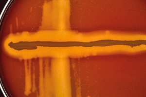
A. pleuropneumoniae is a non-motile, Gram-negative, encapsulated coccobacillus bacteria found in the Pasteurellaceae family. It exhibits β-hemolysis activity, thus explaining its growth on chocolate or blood agar, but must be supplemented with V factor (NAD) in order to facilitate growth for one of its biological variants. As a facultative anaerobic pathogen, A. pleuropneumoniae may need CO2 in order to grow.Depending on the biovar, the bacteria may or may not be positive for urease; both biovars are positive for porphyrin. [2]
Ecology and Pathogenesis
The bacterium rapidly colonizes the host and attaches to the epithelial cells of the tonsils, moving down to the respiratory tract utilizing type IV fimbriae.As the bacteria replicate, they release cytotoxins, hemolysins and the LPS on their outer membranes.The subsequent lysis of macrophages causes a release of lysozymes, which in turn cause the tissue damage seen in porcine pleuropneumonia. Members of the Pasteurellaceae family routinely change the cellular processes of the infected cell.In particular, A. pleuropneumoniae activates the creation of various cytokines such as interleukin 1β (IL-1β), IL-8 and tumor necrosis factor-alpha (TNF-α). IL-8 is itself a chemical signal used to attract neutrophils to the infection site.
The typical presentation of A. pleuropneumoniae in pigs is the characteristic demarcated lesions in the middle, cranial and caudal lobes of the lung. Areas of severe pneumonic growth are dark and consolidated.In the case of chronically infected pigs, pleural adhesions and abscesses are normally found. Histological studies of infected lung tissue will normally showcase lung necrosis, neutrophil infiltration, macrophage and platelet activation and an exudate. Severe hemolysis or hemorrhaging is also present.
Several virulence factors account for the remarkable pathogenesis of Actinobacillus pleuropneumoniae. The more important ones include the production and release of the Apx toxins, the ability to produce a biofilm, its LPS layer, capsule polysaccharides and its ability to survive within an iron-limited environment. Out of these, the most important are its capsule and Apx toxin production.
References
[1] Kokotovic, B., Angen, O., and Bisgaard, M. 2011. “Genetic diversity of Actinobacillus lignieresii isolates from different hosts”. International Journal of Acta Veterinaria Scandinavica. 53(1):6. http://www.actavetscand.com/content/53/1/6 [2] Marsteller, TA; Fenwick B (1999). "Actinobacillus pleuropneumoniae disease and serology". Swine Health and Production 7 (4): 161–165. [3] Auger, E; Deslandes V, Ramjeet M, et al. (2009). "Host-pathogen interactions of Actinobacillus pleuropneumoniae with porcine lung and tracheal epithelial cells". Infection and Immunity 77 (4): 1426–1441. https://www.ncbi.nlm.nih.gov/pubmed/19139196?dopt=Abstract
Author
Page authored by Kejun Shao, student of Mandy Brosnahan, Instructor at the University of Minnesota-Twin Cities, MICB 3301/3303: Biology of Microorganisms.

