Avian Malaria in Hawaiian Birds
]
Plasmodium
While malaria is most commonly thought of in terms of the few species that infect humans, there are several hundreds of haemosporidian parasites that infect a variety of vertebrate hosts [1]
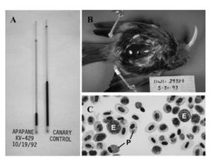
These parasites are commonly split into five genera: Plasmodium, Hepatocystis, Haemoproteus, Parahaemoproteus, and Leucocoytozoon ([2]. Of these genera, species within the Leucocytozoon, Parahaemorptoeus, and Plasmodium genera have been found to infect avian species [3]. Of these, Plasmodium relictum has been devastating the native birds on the island of Hawaii [4].

By Jess Kotnour
At right is a sample image insertion. It works for any image uploaded anywhere to MicrobeWiki.
The insertion code consists of:
Double brackets: [[
Filename: PHIL_1181_lores.jpg
Thumbnail status: |thumb|
Pixel size: |300px|
Placement on page: |right|
Legend/credit: Electron micrograph of the Ebola Zaire virus. This was the first photo ever taken of the virus, on 10/13/1976. By Dr. F.A. Murphy, now at U.C. Davis, then at the CDC.
Closed double brackets: ]]
Other examples:
Bold
Italic
Subscript: H2O
Superscript: Fe3+
Sample citations: [6]
[7]
A citation code consists of a hyperlinked reference within "ref" begin and end codes.
Life Cycle of Plasmodium relictum
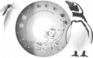
Plasmodium relictum follows the common haemosporidian life cycle. P. relictum initially undergoes a period of asexual reprodcution within mosquito host tissues (LaPointe et al 2012). This initial cycle is followed by subsequent cycles of asexual reproduction within both host tissues and red blood cells, leading to gametocytes within host red blood cells (LaPointe et al 2012). These gametocytes will remain in the midgut, becoming gametes until forming fertile zygotes (LaPointe et al 2012). These zygotes will eventually form into oocysts. Inside the oocysts, asexual reproduction will occur to form sporozoties (Atkinson and Van Riper III YEAR). As they rupture from oocysts, these sporozoties will move into the salivary glands of the mosquito, where they can be transmitted intravenously, intramuscularly, or intraperitoneally to birds (Atkinson and Van Riper III YEAR).
Once inside the avian host, the life cycle continues. The sporozoties will invade the avian host’s reticuloendothelial (macrophages and monocytes) cells where they will develop into mernots (Grilo et al. 2016). Through asexual reproduction, mernots will reproduce into merozites while still inside the host’s reticuloendothelial cells (Grilo et al 2016). When the cell inevitably ruptures, the merozites will be released and will spread throughout the host using the blood stream (Grilo et al 2016). P. relictum continues its development in reticuloendothelial and host tissues (Grilo et al 2016). They most commonly develop in the liver, spleen, and kidneys of the host (Grilo et al 2016). Once inside either the host’s cells or tissue, the merozites will further develop into either erythrocytic mernots or gametocytes (Grilo et al 2016). If the merozite develops into a gametocyte, the development is complete, and it will remain in the host’s red blood cells until a mosquito vector bites the host (Grilo et al 2016). If the host is bitten by a mosquito, the gametocyte will repeat the life cycle in the mosquito.
P. relictum often infects birds most often during the breeding months (Atkinson and Van Riper III year). Breeding season is often during the warmer months of the year, allowing for the development mosquitos and P. relictum (Atkinson and Van Ripper III). These mosquitos are able to take advantage of the breeding season, as birds are often investing more resources into raising their young than their immune systems (Atkinson and Van Ripper III Year). The weekend immune system makes infection by P. relictum much easier (Atkinson and Van Ripper III YEAR).
Include some current research, with at least one figure showing data.

Every point of information REQUIRES CITATION using the citation tool shown above.
Section 2
Include some current research, with at least one figure showing data.

Section 3
Include some current research, with at least one figure showing data.
Section 4
Conclusion
References
- ↑ Martinsen and Perkins 2013. The Diversity of Plasmodium and other Haemosporidians: The Intersections of Taxonomy, Phylogenetics, and Genomics. In: Carlton, J., Perkins, S., Deitsch, editors. Malaria Parasites: Comparative Genomics, Evolution and Molecular Biology. Norfolk: Caister Academic Press. p 1- 15.
- ↑ Martinsen and Perkins 2013. The Diversity of Plasmodium and other Haemosporidians: The Intersections of Taxonomy, Phylogenetics, and Genomics. In: Carlton, J., Perkins, S., Deitsch, editors. Malaria Parasites: Comparative Genomics, Evolution and Molecular Biology. Norfolk: Caister Academic Press. p 1- 15.
- ↑ Martinsen and Perkins 2013. The Diversity of Plasmodium and other Haemosporidians: The Intersections of Taxonomy, Phylogenetics, and Genomics. In: Carlton, J., Perkins, S., Deitsch, editors. Malaria Parasites: Comparative Genomics, Evolution and Molecular Biology. Norfolk: Caister Academic Press. p 1- 15.
- ↑ LaPointe et al.: Ecology and conservation biology of avian malaria. Annals of the New York Academy of Sciences 2012. 1249:211-226
- ↑ LaPointe et al.: Ecology and conservation biology of avian malaria. Annals of the New York Academy of Sciences 2012. 1249:211-226
- ↑ Hodgkin, J. and Partridge, F.A. "Caenorhabditis elegans meets microsporidia: the nematode killers from Paris." 2008. PLoS Biology 6:2634-2637.
- ↑ Bartlett et al.: Oncolytic viruses as therapeutic cancer vaccines. Molecular Cancer 2013 12:103.
- ↑ [https://www.tandfonline.com/doi/full/10.1080/03079457.2016.1149145 Grilo et al.2016. Malaria in penguins—current perceptions. Avian Pathology. 45(4): 393-407. ]
- ↑ Atkinson et al. 2000. Pathogenicity of avian malaria in experimentally infected Hawaii Amakihi. Journal of Wildlife Diseases. 36(2): 197-204
Authored for BIOL 238 Microbiology, taught by Joan Slonczewski, 2018, Kenyon College.
