Bacterial nucleation in pseudomonas syringae: Difference between revisions
From MicrobeWiki, the student-edited microbiology resource
OconnorRyan (talk | contribs) No edit summary |
OconnorRyan (talk | contribs) No edit summary |
||
| Line 3: | Line 3: | ||
=Pseudomonas syringae= | =Pseudomonas syringae= | ||
== | ==Species== | ||
[[Image:Pseudosmonas_syringae_SEM.jpg|thumb|400px|right|[Image 1] | [[Image:Pseudosmonas_syringae_SEM.jpg|thumb|400px|right|[Image 1] | ||
| Line 16: | Line 16: | ||
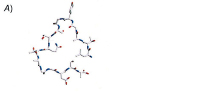
[[Image:Ice_Nucleating_Protein.png|thumb|400px|right|[Image 3] <br><i>Pseudomonas syringae</i> Cross section of modeled INP and a B-helical protein showing a wire frame representation of one loop. Cross section after 100 steps of energy minimization. Source: Graether and Jia, (2001).]] | [[Image:Ice_Nucleating_Protein.png|thumb|400px|right|[Image 3] <br><i>Pseudomonas syringae</i> Cross section of modeled INP and a B-helical protein showing a wire frame representation of one loop. Cross section after 100 steps of energy minimization. Source: Graether and Jia, (2001).]] | ||
== | ==Description== | ||
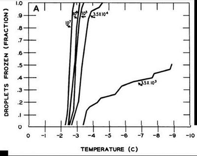
[[Image:Lindow et al.jpg|thumb|400px|right|[Figure 1] <br> Proportions of frozen droplets with respect to temperature at <i>Pseudomonas syringae</i> concentrations of 10<sup>7</sup>, 10<sup>6</sup>, 10<sup>5</sup>, 3.5<sup>4</sup>, and 3.5<sup>3</sup>. Cells were grown on nutrient agar and suspended in sterile water at appropriate concentrations. Source: Lindow <i>et al.</i>, 1982.]] | [[Image:Lindow et al.jpg|thumb|400px|right|[Figure 1] <br> Proportions of frozen droplets with respect to temperature at <i>Pseudomonas syringae</i> concentrations of 10<sup>7</sup>, 10<sup>6</sup>, 10<sup>5</sup>, 3.5<sup>4</sup>, and 3.5<sup>3</sup>. Cells were grown on nutrient agar and suspended in sterile water at appropriate concentrations. Source: Lindow <i>et al.</i>, 1982.]] | ||
Revision as of 02:04, 24 April 2011
Introduction
Pseudomonas syringae
Species
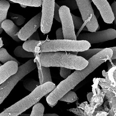
[Image 1]
Pseudomonas syringae shown using SEM. Source: Gordon Vrdoljak, Electron Microscopy Laboratory, U.C. Berkeley [1]
Pseudomonas syringae shown using SEM. Source: Gordon Vrdoljak, Electron Microscopy Laboratory, U.C. Berkeley [1]
Importance
Ice Nucleation Active (INA) Proteins
Description
Structure
Bacterial Effects
Current Research
Conclusion
References
[1] http://genome.jgi-psf.org/psesy/psesy.home.html [2]
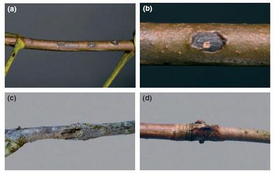
[2] Steele, H., B.E. Laue, G.A. MacAskill, A.J. Hendry, and S. Green. “Analysis of the natural infection of European horse chestnut (Aesculus hippocastanum) by Pseudomonas syringae pv. Aesculi.” Plant Pathology 59: 1005-1013.
[3] Graether, S.P., and Z. Jia. 2001. Modeling Pseudomonas syringae ice-nucleation protein as a B-helical protein. Biophysical Journal 80: 1169-1173.
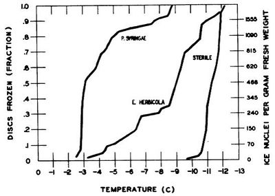
[4] Lindow, S.E., D.C. Arny, and C.D. Upper. 1982. Bacterial ice nucleation: a factor in frost injury to plants. Plant Physiology 70: 1084-1089.
Edited by Ryan O'Connor,student of Joan Slonczewski for BIOL 238 Microbiology, 2011, Kenyon College.
