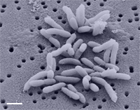
Brevundimonas diminuta
Classification
Bacteria; Proteobacteria; Alphaproteobacteria; Caulobacterales; Caulobacteraceae; Brevundimonas; Brevundimonas diminuta
Brevundimonas diminuta

Description and Significance
Brevundimonas diminuta is non-lactose-fermenting environmental Gram-negative bacilli [1] . B. diminuta and Brevundimonas vesicularis were previously assigned to the genus Pseudomanas. However, the numerical analyses of whole-cell protein patterns, fatty acid compositions, phenotypic characteristics, DNA base ratios, and degrees of DNA relatedness showed that they should be categorized in a separate genus [2] . B. diminuta is commonly used as a test organism for validation of sterilizing-grade membrane filters due to the small size of the bacterium [3] .
Structure, Metabolism, and Life Cycle
Brevundimonas diminuta is actively motile with predominantly a single polar flagellum. One unusual morphological feature is that many of the individuals have flagellum originating from the periphery of the pole rather than the center of the pole [4] . B. diminuta grows very readily in a simple peptone solution at optimal pH around 7 and at optimal temperature around 35°C [4] . The organism does not ferment any carbohydrates and shows no hemolysis activity, and it oxidizes ethanol to acid [4] .
Ecology and Pathogenesis
Brevundimonas diminuta is rarely isolated from environmental specimens (water) and clinical specimens (blood). Even though it is not considered pathogenic, there have been multiple clinical case reports relating this microbe with infections in patients with cancer [1] . All clinical strains that were tested showed that this microbe is intrinsically resistant to fluoroquinolones [1] .
References
1. Han, X., and Andrade, R. 2005. “Brevundimonas diminuta infections and its resistance to fluoroquinolones”. “Journal of Antimicrobial Chemotherapy” 55 (6): 853-859. http://jac.oxfordjournals.org/content/55/6/853.full
2. Segers, P., Vancanneyt, M., Pot, B., Torck, U., Hoste, B., Dewettinck, D., Falsen, E., Kersters, K., and De Vos, P. 1994. “Classification of Pseudomonas diminuta Leifson and Hugh 1954 and Pseudomonas vesicularis Busing, Doll, and Freytag 1953 in Brevundimonas gen. nov. as Brevundimonas diminuta comb. nov. and Brevundimonas vesicularis comb. nov., Respectively”. “International Journal of Systematic and Evolutionary Microbiology”. 44(3): 499-510. http://ijs.sgmjournals.org/content/44/3/499.full.pdf+html
3. Lee, S., Lee, S., and Kim, C. 2002. “Changes in the Cell Size of Brevundimonas diminuta Using Different Growth Agitation Rates”. “PDA Journal of Pharmaceutical Science and Technology”. 56 (2): 99-108. http://journal.pda.org/content/56/2/99.short
4. Leifson, E., and Hugh, R. 1954. “A New Type of Polar Monotrichous Flagellation.” “Microbiology.” 10: 68-70. http://mic.sgmjournals.org/content/10/1/68.full.pdf+html
Author
Page authored by Heekyung Lim, student of Mandy Brosnahan, Instructor at the University of Minnesota-Twin Cities, MICB 3301/3303: Biology of Microorganisms.
