File:Mmi 867 f7.gif
From MicrobeWiki, the student-edited microbiology resource
Mmi_867_f7.gif (150 × 191 pixels, file size: 16 KB, MIME type: image/gif)
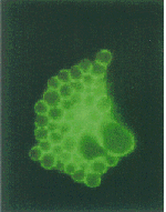
Immunofluorescence microscopy of C. psittaci GPIC-infected HeLa cells at 30 h PI. The α-IncA-stained cells were fixed with methanol (A); the α-IncC-stained cells were fixed with periodate–lysine–paraformaldehyde (PLP) fixative (B) and α-IncB-stained cells fixed in methanol (C). The immunofluorescent images show staining of the inclusion membrane, but no chlamydial staining with any antisera. Bar is 10 μm. [copyright Bannantine, J. P]
File history
Click on a date/time to view the file as it appeared at that time.
| Date/Time | Thumbnail | Dimensions | User | Comment | |
|---|---|---|---|---|---|
| current | 05:54, 29 August 2007 |  | 150 × 191 (16 KB) | Coho (talk | contribs) | Immunofluorescence microscopy of C. psittaci GPIC-infected HeLa cells at 30 h PI. |
| 05:40, 29 August 2007 |  | 914 × 422 (212 KB) | Coho (talk | contribs) | Immunofluorescence microscopy of C. psittaci GPIC-infected HeLa cells at 30 h PI. The α-IncA-stained cells were fixed with methanol (A); the α-IncC-stained cells were fixed with periodate–lysine–paraformaldehyde (PLP) fixative (B) and α-IncB-staine |
You cannot overwrite this file.
File usage
The following page uses this file:

