Herpesviridae and Their Role in Uveitis: Difference between revisions
No edit summary |
No edit summary |
||
| Line 32: | Line 32: | ||
[[File:Cytomegalovirus_retinitis.jpg|200px|thumb|left|Image showing what cytomegalovirus looks like. From Hudson HL, Boyer DS, Martin DF, et al: Viral posterior uveitis. In Yanoff M, Duker JS [eds]: Ophthalmology, London, Mosby, 1999. http://clinicalgate.com/uveitis/]] <br> | [[File:Cytomegalovirus_retinitis.jpg|200px|thumb|left|Image showing what cytomegalovirus looks like. From Hudson HL, Boyer DS, Martin DF, et al: Viral posterior uveitis. In Yanoff M, Duker JS [eds]: Ophthalmology, London, Mosby, 1999. http://clinicalgate.com/uveitis/]] <br> | ||
<br> | <br> | ||
[[File:Keratic_precipitates_in_anterior_uveitis.jpg|200px|thumb|left|Image showing keratic precipitates in uveitis. From Forster DJ: General approach to the uveitis patient and treatment strategies. In Yanoff M, Duker JS [eds]: Ophthalmology, London, Mosby, 1999. http://clinicalgate.com/uveitis/]] | [[File:Keratic_precipitates_in_anterior_uveitis.jpg|200px|thumb|left|Image showing keratic precipitates in uveitis. From Forster DJ: General approach to the uveitis patient and treatment strategies. In Yanoff M, Duker JS [eds]: Ophthalmology, London, Mosby, 1999. http://clinicalgate.com/uveitis/]] <br> | ||
<br> | |||
Herpesviridae, which is most commonly known as herpes, is a family in the order Herpesvirales, that is made up of DNA viruses known for causing diseases in humans and other animals. There are many viruses in this family, more than 130 [1], and there are 3 subfamilies: Alphaherpesvirinae, Betaherpesvirinae, and Gammaherpesvirinae. Out of all Herpesviridae, only eight use humans as their primary host [2]. In the Alphaherpesvirinae group, the viruses are the herpes simplex virus 1, herpes simplex virus 2, and varicella-zoster virus. In the Betaherpesvirinae group, the viruses are the cytomegalovirus, Human herpesvirus-6, and Human herpesvirus-7. In the Gammaherpesvirinae group, the viruses are the Epstein-Barr virus and Kaposi’s sarcoma herpes (or Human herpesvirus-8). The Alphaherpesvirinae virus, the most common Herpesviridae subfamily, have a short replication cycle, that takes about 18 hours. They have a wide range of hosts, and destroy cells efficiently. The Betaherpesvirinae virus replication cycle is long, with a somewhat narrow host range. When they infect cells it results in the cells becoming enlarged (called cytomegalo). The Gammaherpesvirinae virus also has a very narrow host range, and they are specific for only T or B lymphocytes [3, 8]. <br> | |||
<br> | |||
Of the eight herpesviridae capable of infecting humans, five of them are incredibly common. Herpes simplex virus 1 and herpes simplex virus 2 are the cause of genital and orolabial herpes, cytomegalovirus causes mononucleosis and pneumonias, Epstein-Barr virus causes mononucleosis, and the varicella-zoster virus is responsible for both shingles and chicken pox. Of these common viruses, herpes simplex virus, varicella-zoster virus, and cytomegalovirus are each capable of causing acute, recurrent, and chronic uveitis [4]. All herpesviridae viruses can be latent or lytic. Many people who are infected are unaware because they don’t exhibit any symptoms. <br> | |||
<br> | |||
Uveitis: (figure 1: diagram of the eye) (more in here as overview) (can compare anterior to intermediate to posterior to pan: Anteriror uveitis is the most prevalent form of it) | |||
Uveitis is a form of inflammation in the eye. Specifically, it is the inflammation of the uvea, which is a pigmented layer in the eye that is composed of the iris, ciliary body, and choroid. Uveitis can be in either one eye or both eyes, and it can also involve other parts of the eye such as the cornea, sclera, retina, vitreous body, and/or the optic nerve. It is considered an ophthalmic emergency and it needs to be examined by an optometrist or ophthalmologist, followed by treatment in order to control the inflammation. If left untreated, it will often lead to blindness [5]. Uveitis can be described more specifically based on where in the eye it occurs. There is anterior uveitis, intermediate uveitis, posterior uveitis, and panuveitis uveitis. <br> | |||
<br> | |||
==Section 2== | ==Section 2== | ||
Include some current research, with at least one figure showing data.<br> | Include some current research, with at least one figure showing data.<br> | ||
<br> | <br> | ||
Structure (and genome) (Figure 2: picture of structure) | |||
All Herpesviridae virions have the same unique structure made up of four elements or layers, which is how viruses come to be classified as a Herpesviridae [3]. These are the core, capsid, tegument, and envelope. Its structure is responsible for keeping the virus’s genome safe, as well as enabling the virus to infect the correct target cell. Within the core, there is a molecule of double-stranded DNA. Virus DNA in the Herpesviridae family generally have about 120 to 250 kb pairs. The virus core is inside of an icosahedral capsid, which usually has a diameter of about 100 nm that is made up of 162 capsomeres. Herpes simplex virus capsids are made up of five conserved proteins. These proteins are pUL19, that is responsible for forming all of the capsids, pUL18 and pUL38, that form triplexes so that adjacent capsomeres can be interacted with and stabilized, pUL35, that is responsible for covering hexons, and pUL6, that is responsible for forming a portal [7]. Depending on the type of Herpesviridae virion, there may be other cellular proteins there such as actin, tubulin, heat shock proteins, or annexin [7]. The tegument, which coats this capsid, is a layer of proteins that has viral mRNAs along with viral proteins in it. Surrounding the tegument is a lipid bilayer envelope that contains glycoproteins, lipids, and polyamines in it [3, 7]. | |||
The DNA Herpesviridae has a unique genome sequence arrangement. <br> | |||
<br> | |||
==Section 3== | ==Section 3== | ||
Revision as of 03:34, 29 April 2016
Section
By Chris Link
At right is a sample image insertion. It works for any image uploaded anywhere to MicrobeWiki.
The insertion code consists of:
Double brackets: [[
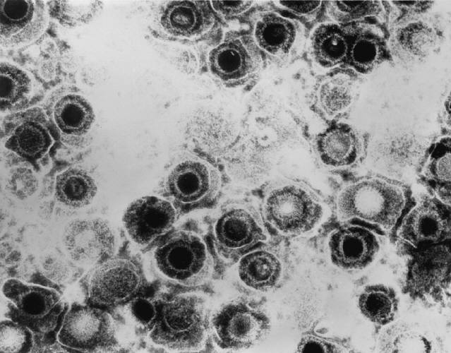
Filename: Transmission_electron_micrograph_of_herpes_simplex_virus.jpg
Thumbnail status: |thumb|
Pixel size: |300px|
Placement on page: |right|
Legend/credit: Transmission electron micrograph of herpes simplex virus. Taken by Erskine Palmer - http://phil.cdc.gov/PHIL_Images/08301998/00014/B82-0474_lores.jpg
Closed double brackets: ]]
Other examples:
Bold
Italic
Subscript: H2O
Superscript: Fe3+
Introduce the topic of your paper. What is your research question? What experiments have addressed your question? Applications for medicine and/or environment?
Sample citations: [1]
[2]
A citation code consists of a hyperlinked reference within "ref" begin and end codes.
[3]
Section 1
Include some current research, with at least one figure showing data.
Every point of information REQUIRES CITATION using the citation tool shown above.


Herpesviridae, which is most commonly known as herpes, is a family in the order Herpesvirales, that is made up of DNA viruses known for causing diseases in humans and other animals. There are many viruses in this family, more than 130 [1], and there are 3 subfamilies: Alphaherpesvirinae, Betaherpesvirinae, and Gammaherpesvirinae. Out of all Herpesviridae, only eight use humans as their primary host [2]. In the Alphaherpesvirinae group, the viruses are the herpes simplex virus 1, herpes simplex virus 2, and varicella-zoster virus. In the Betaherpesvirinae group, the viruses are the cytomegalovirus, Human herpesvirus-6, and Human herpesvirus-7. In the Gammaherpesvirinae group, the viruses are the Epstein-Barr virus and Kaposi’s sarcoma herpes (or Human herpesvirus-8). The Alphaherpesvirinae virus, the most common Herpesviridae subfamily, have a short replication cycle, that takes about 18 hours. They have a wide range of hosts, and destroy cells efficiently. The Betaherpesvirinae virus replication cycle is long, with a somewhat narrow host range. When they infect cells it results in the cells becoming enlarged (called cytomegalo). The Gammaherpesvirinae virus also has a very narrow host range, and they are specific for only T or B lymphocytes [3, 8].
Of the eight herpesviridae capable of infecting humans, five of them are incredibly common. Herpes simplex virus 1 and herpes simplex virus 2 are the cause of genital and orolabial herpes, cytomegalovirus causes mononucleosis and pneumonias, Epstein-Barr virus causes mononucleosis, and the varicella-zoster virus is responsible for both shingles and chicken pox. Of these common viruses, herpes simplex virus, varicella-zoster virus, and cytomegalovirus are each capable of causing acute, recurrent, and chronic uveitis [4]. All herpesviridae viruses can be latent or lytic. Many people who are infected are unaware because they don’t exhibit any symptoms.
Uveitis: (figure 1: diagram of the eye) (more in here as overview) (can compare anterior to intermediate to posterior to pan: Anteriror uveitis is the most prevalent form of it)
Uveitis is a form of inflammation in the eye. Specifically, it is the inflammation of the uvea, which is a pigmented layer in the eye that is composed of the iris, ciliary body, and choroid. Uveitis can be in either one eye or both eyes, and it can also involve other parts of the eye such as the cornea, sclera, retina, vitreous body, and/or the optic nerve. It is considered an ophthalmic emergency and it needs to be examined by an optometrist or ophthalmologist, followed by treatment in order to control the inflammation. If left untreated, it will often lead to blindness [5]. Uveitis can be described more specifically based on where in the eye it occurs. There is anterior uveitis, intermediate uveitis, posterior uveitis, and panuveitis uveitis.
Section 2
Include some current research, with at least one figure showing data.
Structure (and genome) (Figure 2: picture of structure)
All Herpesviridae virions have the same unique structure made up of four elements or layers, which is how viruses come to be classified as a Herpesviridae [3]. These are the core, capsid, tegument, and envelope. Its structure is responsible for keeping the virus’s genome safe, as well as enabling the virus to infect the correct target cell. Within the core, there is a molecule of double-stranded DNA. Virus DNA in the Herpesviridae family generally have about 120 to 250 kb pairs. The virus core is inside of an icosahedral capsid, which usually has a diameter of about 100 nm that is made up of 162 capsomeres. Herpes simplex virus capsids are made up of five conserved proteins. These proteins are pUL19, that is responsible for forming all of the capsids, pUL18 and pUL38, that form triplexes so that adjacent capsomeres can be interacted with and stabilized, pUL35, that is responsible for covering hexons, and pUL6, that is responsible for forming a portal [7]. Depending on the type of Herpesviridae virion, there may be other cellular proteins there such as actin, tubulin, heat shock proteins, or annexin [7]. The tegument, which coats this capsid, is a layer of proteins that has viral mRNAs along with viral proteins in it. Surrounding the tegument is a lipid bilayer envelope that contains glycoproteins, lipids, and polyamines in it [3, 7].
The DNA Herpesviridae has a unique genome sequence arrangement.
Section 3
Include some current research, with at least one figure showing data.
Section 4
Conclusion
References
- ↑ Hodgkin, J. and Partridge, F.A. "Caenorhabditis elegans meets microsporidia: the nematode killers from Paris." 2008. PLoS Biology 6:2634-2637.
- ↑ Bartlett et al.: Oncolytic viruses as therapeutic cancer vaccines. Molecular Cancer 2013 12:103.
- ↑ Errera M.H., Goldschmidt, P., Batellier, L., Degorge S., Héron, E., Laroche, L., Sahel, J.A., Westcott, M., and Chaumeil, C."Real-time polymerase chain reaction and intraocular antibody production for the diagnosis of viral versus toxoplasmic infectious posterior uveitis." 2011. Graefe's Archive for Clinical and Experimental Ophthalmology. V 249 (12): 1837-1846.
Authored for BIOL 238 Microbiology, taught by Joan Slonczewski, 2016, Kenyon College.
