Herpesviridae and Their Role in Uveitis
Section
By Chris Link
At right is a sample image insertion. It works for any image uploaded anywhere to MicrobeWiki.
The insertion code consists of:
Double brackets: [[
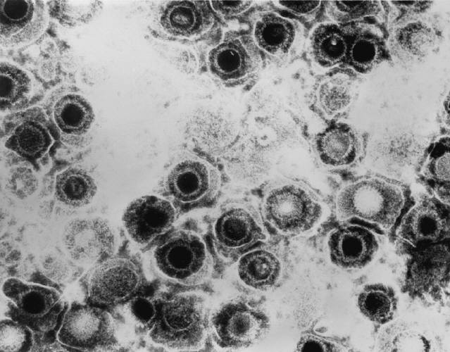
Filename: Transmission_electron_micrograph_of_herpes_simplex_virus.jpg
Thumbnail status: |thumb|
Pixel size: |300px|
Placement on page: |right|
Legend/credit: Transmission electron micrograph of herpes simplex virus. Taken by Erskine Palmer - http://phil.cdc.gov/PHIL_Images/08301998/00014/B82-0474_lores.jpg
Closed double brackets: ]]
Other examples:
Bold
Italic
Subscript: H2O
Superscript: Fe3+
Introduce the topic of your paper. What is your research question? What experiments have addressed your question? Applications for medicine and/or environment?
Sample citations: [1]
[2]
A citation code consists of a hyperlinked reference within "ref" begin and end codes.
[3]
Introduction
Include some current research, with at least one figure showing data.
Every point of information REQUIRES CITATION using the citation tool shown above.


Herpesviridae, which is most commonly known as herpes, is a family in the order Herpesvirales, that is made up of DNA viruses known for causing diseases in humans and other animals. There are many viruses in this family, more than 130 [1], and there are 3 subfamilies: Alphaherpesvirinae, Betaherpesvirinae, and Gammaherpesvirinae. Out of all Herpesviridae, only eight use humans as their primary host [2]. In the Alphaherpesvirinae group, the viruses are the herpes simplex virus 1, herpes simplex virus 2, and varicella-zoster virus. In the Betaherpesvirinae group, the viruses are the cytomegalovirus, Human herpesvirus-6, and Human herpesvirus-7. In the Gammaherpesvirinae group, the viruses are the Epstein-Barr virus and Kaposi’s sarcoma herpes (or Human herpesvirus-8). The Alphaherpesvirinae virus, the most common Herpesviridae subfamily, have a short replication cycle, that takes about 18 hours. They have a wide range of hosts, and destroy cells efficiently. The Betaherpesvirinae virus replication cycle is long, with a somewhat narrow host range. When they infect cells it results in the cells becoming enlarged (called cytomegalo). The Gammaherpesvirinae virus also has a very narrow host range, and they are specific for only T or B lymphocytes [3, 8].
Of the eight herpesviridae capable of infecting humans, five of them are incredibly common. Herpes simplex virus 1 and herpes simplex virus 2 are the cause of genital and orolabial herpes, cytomegalovirus causes mononucleosis and pneumonias, Epstein-Barr virus causes mononucleosis, and the varicella-zoster virus is responsible for both shingles and chicken pox. Of these common viruses, herpes simplex virus, varicella-zoster virus, and cytomegalovirus are each capable of causing acute, recurrent, and chronic uveitis [4]. All herpesviridae viruses can be latent or lytic. Many people who are infected are unaware because they don’t exhibit any symptoms.
Uveitis:
Uveitis is a form of inflammation in the eye. Specifically, it is the inflammation of the uvea, which is a pigmented layer in the eye that is composed of the iris, ciliary body, and choroid. Uveitis can be in either one eye or both eyes, and it can also involve other parts of the eye such as the cornea, sclera, retina, vitreous body, and/or the optic nerve. It is considered an ophthalmic emergency and it needs to be examined by an optometrist or ophthalmologist, followed by treatment in order to control the inflammation. If left untreated, it will often lead to blindness [5]. Uveitis can be described more specifically based on where in the eye it occurs. There is anterior uveitis (the inflammation of iris, or of both the iris and ciliary body), intermediate uveitis (inflammation of the cilliary), posterior uveitis (inflammation of the choroid), and panuveitis uveitis (inflammation in all areas of the uvea).
Herpesviridae Structure
Include some current research, with at least one figure showing data.
All Herpesviridae virions have the same unique structure made up of four elements or layers, which is how viruses come to be classified as a Herpesviridae [3]. These are the core, capsid, tegument, and envelope. Its structure is responsible for keeping the virus’s genome safe, as well as enabling the virus to infect the correct target cell. Within the core, there is a molecule of double-stranded DNA. Virus DNA in the Herpesviridae family generally have about 120 to 250 kb pairs. The virus core is inside of an icosahedral capsid, which usually has a diameter of about 100 nm that is made up of 162 capsomeres. Herpes simplex virus capsids are made up of five conserved proteins. These proteins are pUL19, that is responsible for forming all of the capsids, pUL18 and pUL38, that form triplexes so that adjacent capsomeres can be interacted with and stabilized, pUL35, that is responsible for covering hexons, and pUL6, that is responsible for forming a portal [7]. Depending on the type of Herpesviridae virion, there may be other cellular proteins there such as actin, tubulin, heat shock proteins, or annexin [7]. The tegument, which coats this capsid, is a layer of proteins that has viral mRNAs along with viral proteins in it. Surrounding the tegument is a lipid bilayer envelope that contains glycoproteins, lipids, and polyamines in it [3, 7].
Section 3
Include some current research, with at least one figure showing data.
Section 4
Diagnosis & treatment
Since uveitis has the potential to cause severe eyesight loss or blindness, it is very important that it gets diagnosed correctly, including the cause behind the uveitis. If something is diagnosed incorrectly, the treatments will be wrong, which can possibly lead to other potential problems. Diagnosis can sometimes be a problem, especially with inexperienced ophthalmologists, because some of the disease’s signs can be very hard to identify. Even if the disease is seen to be as a result of a herpesviridae virus, it is vital to know which virus it was. Therefore, it must be determined if it was a herpes simplex virus, a varicella-zoster virus, or a cytomegalovirus virus.
Uveitis associated with herpes simplex virus is most common in people younger than 60 years old, however, it is possible to at any age. In most cases, this occurs one eye only, although it is possible in both eyes. Also, this condition is usually linked to acute ocular hypertension, or raised intraocular pressure in the eye, but not always. Sometimes, there will be iris atrophy, which is an out of place pupil, that will strongly suggest the cause of the uveitis to be herpes simplex virus. Also, a history of herpes simplex virus on the subject’s lips or genitals will help point to an answer, but that is not absolutely necessary. Some other symptoms to look for in a subject include large and central keratic precipitates (this is an inflammatory cellular deposit that would not be a worry if they were more evenly distributed on the corneal endothelium), dilated pupils, and a defect in iris transillumination [4]. Individually, none of the symptoms will result in a conclusion of herpes simplex virus, but when put together, a result can be found. Effective treatments for uveitis associated with herpes simplex virus have been found. It is recommended to take systemic antivirals as the primary treatment, as they will help to protect the cornea in the eye along with treating the uveitis. It has also been observed that if a topical antiviral is used for an extended period of time, the infected person is at risk for the development of keratopathy, which is a corneal disease that is caused by calcium appearing on the central cornea, which can lead to calcification. In addition, using topical corticosteroids also has a potential to cause some problems, mainly because of they are extremely hard for those infected with herpes to stop taking, resulting in a long and slow process in order to taper off of them [4].
(figure 3: varicella zoster virus in eye)Uveitis associated with varicella-zoster virus is most common in people older than 60 years old, but it possible at any age. One identifying characteristic of this virus causing uveitis is what looks like a little bit of string in the cornea of the infected person. This is caused by a special form of pseudodendrite that has cells amassed together in the center with no ulcerations. Varicella-zoster viruses in uveitis are also linked to greatly lessened corneal feelings, which essentially the reason for the blink reflex. Additionally, the symptoms seen in herpes simplex virus associated with uveitis can also be seen in varicella-zoster virus associated uveitis. However, the two can be easily differentiated because those infected with the varicella-zoster virus almost always has a history of dermatitis/shingles, or inflammation of the skin, caused by the virus, and it always occurs on the same side of the body. Treatment for this virus is similar to the herpes simplex virus treatment, so systemic antivirals are taken, except that the dose is doubled. To prevent the varicella-zoster virus, physicians use Varivax for chickenpox and Zostavax for shingles [4].
Uveitis has just relatively recently started to be associated with the cytomegalovirus virus. This virus causes uveitis much less frequently than the herpes simplex virus or the varicella-zoster virus. Although rare in both people over and under 60 years old, it is more prevalent in those under 60. The most identifying characteristic of this virus being associated with uveitis is a nummular keratic precipitate. This means that there are lesions that appear in a coin shape in the stroma. They appear as an immune reaction to viral antigens [6]. Since the cytomegalovirus is so rare, it is usually only considered after a patient with symptoms of uveitis from other Herpesveridae viruses has been treated with corticosteroids as well as systemic antivirals. The cytomegalovirus virus in association with uveitis can usually be treated with oral antiviral medication called ganciclovir, but there is possibility for relapse after the treatments are ended [4]. Because the cytomegalovirus in uveitis is a relatively new finding, more work on this virus’s association with uveitis needs to be done. This is especially because its treatment, ganciclovir, has toxicity problems, and it remains unknown what effects may occur after extended use of it [4].
Section 4
Herpesviridae Cycle
The Herpesviridae virus needs multiple steps in order replicate. Because the Herpesviridae family is made up of viruses that all share the same structure, the cycle they all go through are similar. By explaining the cycle that the herpes simplex virus goes through, an understanding of all Herpesviridae virus cycles can be gained. First, the virion must attach to the host cell. The virion has multiple envelope proteins that are capable of binding to one of several receptors on the target cell surface. After attachment, the virion uses the glycoproteins in its envelope to fuse its membrane with the membrane of the host. After fusion is complete, the virus’s entire capsid is released into the host cytoplasm. This full herpes capsid will then travel down what can be considered a ‘scaffold’ of microtubules, where it will eventually reach the nuclear membrane where a pore is located. As this happens, there is a virion host shutoff factor, called Vhs, that will degrade the mRNA of the host, causing the host to cease its protein synthesis [8]. Then, the herpes capsid will inject its DNA through the nuclear pore into the nucleus of the host cell. This is possible through the high pressure in the capsid, caused by the double stranded DNA being in the relatively small capsid. Once inside the nucleus, the viral DNA forms a circle similar to a plasmid. The viral DNA in the nucleus is transcribed to mRNA by the RNA polymerase ll of the host. Then, the viral genome can either start the infection cycle or go for latent infection. For latent infection, mRNA is used to encode LAT proteins, which are responsible for maintaining the virus’s latency, which last for decades until it switches to being lytic. On the other hand, to start the infection cycle, lytic mRNA leaves the nucleus in order to be translated by ribosomes. After translation, the proteins, ready to be put into capsids, re-enter the nucleus. In order to create progeny genomes, the viral DNA still in the nucleus is replicated by viral enzymes along with some enzymes provided by the host cell, and the rolling-circle method of DNA replication is used to create concatemeric DNA. This new DNA will result in some late-stage mRNA, and it will leave the nucleus to be translated, which will result in some envelope proteins. Once they have been translated, the envelope proteins move back through the endoplasmic reticulum and back into the nuclear membrane. Then, the additional late proteins come back to the nucleus, where they get put inside capsids that have DNA in them that has been cleaved into linear chromosomes. After that, an envelope, which was created by the outer nuclear membrane, is formed around the capsid. Then, the virion is sent through the endoplasmic reticulum (where they get a second envelope) as they continue on their way to the Golgi. Finally, as they reach the cell membrane of the host cell, the second envelope fuses with the cell membrane, causing the host cell to release the virions through exocytosis. If many virions are released quickly, the host cells are killed, leading to the sores that are seen when someone has herpes.
Conclusion
References
- ↑ Hodgkin, J. and Partridge, F.A. "Caenorhabditis elegans meets microsporidia: the nematode killers from Paris." 2008. PLoS Biology 6:2634-2637.
- ↑ Bartlett et al.: Oncolytic viruses as therapeutic cancer vaccines. Molecular Cancer 2013 12:103.
- ↑ Errera M.H., Goldschmidt, P., Batellier, L., Degorge S., Héron, E., Laroche, L., Sahel, J.A., Westcott, M., and Chaumeil, C."Real-time polymerase chain reaction and intraocular antibody production for the diagnosis of viral versus toxoplasmic infectious posterior uveitis." 2011. Graefe's Archive for Clinical and Experimental Ophthalmology. V 249 (12): 1837-1846.
1. Brown, J.C., William W. Newcomb (2011). Herpesvirus Capsid Assembly: Insights from Structural Analysis. Current Opinion in Virology 1 (2): 142–149. doi:10.1016/j.coviro.2011.06.003. PMC 3171831. PMID 21927635.
2. Virus Pathogen Resource. About the Herpesviridae family. 2016. Web. http://www.viprbrc.org/brc/aboutPathogen.spg?decorator=herpes
3. Whitley RJ. Herpesviruses. In: Baron S, editor. Medical Microbiology. 4th edition. Galveston (TX): University of Texas Medical Branch at Galveston; 1996. Chapter 68. Available from: http://www.ncbi.nlm.nih.gov/books/NBK8157/
4. Doran, Marianne. 2009. Understanding and Treating Viral Anterior Uveitis. American Academy of Ophthalmology. http://www.aao.org/eyenet/article/viral-anterior-uveitis?september-2009
5. National Eye Institute. Facts about uveitis. 2011. Web. https://nei.nih.gov/health/uveitis/uveitis
6. American Academy of Ophthalmology. Herpes Zoster Ophthalmicus. http://www.aao.org/focalpointssnippetdetail.aspx?id=8367b620-245c-4ebf-89e7-eca0c8d35aa3
7. Mettenleiter, T. C. 2002. Herpesvirus Assembly and Egress. Journal of Virology, 76(4), 1537–1547. http://doi.org/10.1128/JVI.76.4.1537-1547.2002
8. Taddeo, B., & Roizman, B. 2006. The Virion Host Shutoff Protein (UL41) of Herpes Simplex Virus 1 Is an Endoribonuclease with a Substrate Specificity Similar to That of RNase A. Journal of Virology, 80(18), 9341–9345. http://doi.org/10.1128/JVI.01008-06
9. Los Alamos National Laboratory. Herpesvirus Family: Herpesviridae. 2001. Web. http://stdgen.northwestern.edu/stdgen/bacteria/hhv2/herpes.html#sub
10. iKnowledge. 2015. Uveitis. Web. http://clinicalgate.com/uveitis/
11. The ocular immunology and uveitis foundation. 2008. Pars Planitis. Web. http://www.uveitis.org/patients/support/parsplantis-org
12. Yao, Lijing, Agha Hassan Feroze. Herpes Simplex Uveitis. Web. http://www.uveitis.org/docs/dm/herpes_simplex_uveitis_scleritis.pdf
Authored for BIOL 238 Microbiology, taught by Joan Slonczewski, 2016, Kenyon College.
