Nosema ceranae: Difference between revisions
Laceyberry (talk | contribs) (Created page with "WIKI IN PROGRESS Ex. ''[[]]'' ==Characteristics of the symbiont/pathogen== What kind of microbe is it (eg Cell morphology, shape, phylogenetic classification)? Is its genome seq...") |
No edit summary |
||
| (60 intermediate revisions by 2 users not shown) | |||
| Line 1: | Line 1: | ||
[[Category:Short pages]] | |||
WIKI IN PROGRESS | WIKI IN PROGRESS | ||
==Characteristics of the symbiont/pathogen== | ==Characteristics of the symbiont/pathogen== | ||
[[File:NC.jpg|thumb|''Nosema ceranae'' [http://www.kimesapiary.com/images/NosemaCeranae.jpg]]] | |||
''Nosema ceranae'', a microsporidium fungi, is part a member of the Nosematidae family. ''N. ceranae'' is a spore forming, rod or oval shaped microbe that measures approximately 3.9-5.3 µm in length and 2.0-2.5 µm in width. ''Nosema ceranae'' has three developmental stages: Meronts, which is the earliest stage, and during this stage the plasma membrane of the microbe makes direct contact with the cytoplasm of the host. During sporont stage the microbe becomes elongated and oval and consists of a dense cytoplasm, yet there is no distinct internal structures. The third stage is the Sporoblast stage, the microbe is smaller during this stage than the sporont stage, and has a distinct cell wall as well as two nuclei [2]. A complete sequencing of ''N. ceranae'''s genome revealed a size of 7.86 MB with a strong AT bias [3]. | |||
==Characteristics of the host== | ==Characteristics of the host== | ||
[[File:AM.jpg|thumb|''Nosema ceranae'' [9]]] | |||
[[File: | |||
The symbiont relationship with ''N. ceranae'' and the honey bee originated in the Asian honey bee ''Apis cerana'', but has since then switched host to infect the European honey bee ''Apis mellifera''. ''Nosema ceranae'' infects most of the honey bees found in Europe and North America [3]. | |||
==Host-Symbiont Interaction == | ==Host-Symbiont Interaction == | ||
''N. ceranae'' and the honeybee have a obligate parasitic relationship, meaning that the ''N. ceranae'' benefits while the honey bee is harmed throughout the process. ''N. ceranae'' forms spores which are then ingested by the honey bee through water or food. These spores then invade the gut epithelium immediately [3]. However, it is not until day six or seven that symptoms are visible such as ecoming less active, immobile, having an increase in appetite, and usually by day eight mortality has occurred [6,8]. The spores rapidly multiply in the gut and then are excreted and the spores are transferred to the other honeybees that live in the same colony through the cleaning and feeding activities [2]. ''Nosema ceranae'' decrease the ability of the honey bee to obtain nutrients from the environment which ultimately shortens their lifespan. ''Nosema ceranae'' can effect the overall colony growth and hinder winter survival [3]. | |||
==Molecular Insights into the Symbiosis== | ==Molecular Insights into the Symbiosis== | ||
There is no specific gene known to be disrupted or the cause of this infection. ''N. ceranae'' produces spores which then invade the cell, however molecular techniques such as Polymerase Chain Reaction (PCR), which amplifies a certain gene or part of a gene, allows us to see how the host cell is being effected. For example by using PCR and amplifying the 16S rRNA gene researchers are able to detect coinfection of the host by two different species [7]. | |||
==Ecological and Evolutionary Aspects == | ==Ecological and Evolutionary Aspects == | ||
This symbiont relationship between ''Nosema ceranae'' and ''Apis mellifera'' is a recent discovery, and new studies are continually done to uncover more about this parasitic relationship. Originally ''Nosema ceranae'' infected the Asian honey bee (''Apis cerana''), but has since then swtiched to the European honey bee (''Apis mellifera''). It is unknown when and how this switch occurred, and researchers are unsure if the predominant parasite of honey bees in Europe and North America still remains ''N. apis'' or if ''N. ceranae'' has taken over [3]. The biological cycle of ''Nosema ceranae'' relies on the temperature. ''Nosema ceranae'' effects more cells when kept at a constant temperature, but is a better adapter to conditions the other species such as ''Nosema apis''. ''N. ceranae'' unlike ''N. apis'' can infect their host in all four seasons, while ''N. apis'' usually only infects their host in the milder seasons such as spring and autumn [5]. | |||
==Recent Discoveries== | ==Recent Discoveries== | ||
[[File:NC2.jpg|thumb|''Nosema ceranae'' [4]]] | |||
In past studies it was thought that ''N. ceranae'' was becoming the more prevalent parasite to the Honey bee, however recent studies have shown otherwise. ''N. apis'' in a 5 year long study showed to grow in greater quantity then ''N. ceranae'' except in one colony. However it was also found that ''N. ceranae'' is a more general infection, after displaying the ability to grow in other tissues then just the gut. In this five year long study ''N. ceranae'' was observed to grow in not only the gut, but the hypopharyneal glands, salivary glands, malpighian tubes, fat bodies, and the brain. This could account for the hypothesis that ''N. ceranae'' causes more deaths in honey bee colonies then ''N. apis''. It was also found that ''N. ceranae'' during the germination process when exposed to low temperature decreased to less the 10 percent. The remaining spores that where able to germinate were unusually short and most likely did not complete the true germination process [4]. | |||
==References== | ==References== | ||
[Sample reference] [[http://www.plosone.org/article/info%3Adoi%2F10.1371%2Fjournal.pone.0015830] Seemanapalli SV, Xu Q, McShan K, Liang FT. 2010. Outer surface protein C is a dissemination-facilitating factor of ''Borrelia burgdorferi'' during mammalian | [Sample reference] [[http://www.plosone.org/article/info%3Adoi%2F10.1371%2Fjournal.pone.0015830] Seemanapalli SV, Xu Q, McShan K, Liang FT. 2010. Outer surface protein C is a dissemination-facilitating factor of ''Borrelia burgdorferi'' during mammalian | ||
infection. PLoS One 5:e15830.] | infection. PLoS One 5:e15830.] | ||
[1] http://www.diark.org/img/species_pict/Nosema_ceranae_BRL01 | |||
[2]Chen,Y., Evans,J., Murphy,C., Gutell,R., Zuker,M., Gundensen-Rindal,D and Pettis,J. 2009. Morphological, molecular, and phylogenetic characterization of ''Nosema ceranae'', a microsporidian parasite isolated from the European honey bee, ''Apis mellifera''. Journal of Eukaryotic Microbiology 56: 142-147. | |||
[3]Cornman, R., Chen, Y., Schatz,M., Street,C., Zhao,Y., Desany, B., Egholm, M., Hutchison, S., Pettis, J., Lipkin and W., Eva, J. 2009. Genomic Analyses of the Microsporidian ''Nosema ceranae'', an Emergent Pathogen of Honey Bees. PLoS Pathogens 5(6): e1000466. | |||
[4]Gisder S., Hedtke K., Möckel N., Frielitz MC, Linde A, and Genersch E. 2010. Five-year cohort study of Nosema spp. in Germany: does climate shape virulence and assertiveness of Nosema ceranae? Applied Environmental Microbiology 76(9): 3032-3038. | |||
[5]Hernandez, R., Meana,A., Palencia,P., Marín, P., Botías,C., Bailon, E., Barrios, L. and Higes, M., 2009. Effect of Temperature on the Biotic Potential of Honeybee Microsporidia. Applied and Enviromental Microbiology 75(8):2554. | |||
[6]Higes, M., Garcia-Palencia, P., Martin-Hernandez, R. and Meana, A. 2006. Experimental infection of Apis mellifera honeybees with Nosema ceranae (Microsporidia). Journal of Invertbrate Pathology 94: 211-217. | |||
[7]Higes,M., Salvador, A., Garrido-Bailón, E., Martín-Hernández,R., Meana, A. and Prieto, L. 2007. Outcome of Colonization of Nosema ceranaeby Apis mellifera. Applied and Environmental Microbiology 73(20):6331. | |||
[8]Mayack, C. and Naug, D. 2009 Energetic stress in the honeybee Apis mellifera from Nosema ceranae infection. Journal of Invertebrate Pathology 100:185–188. | |||
[9]http://tolweb.org/tree/ToLimages/Apis_mellifera2116051.250a.jpg | |||
Edited by [Lacey Berry], student of [mailto:glim@rmc.edu Grace Lim-Fong] | Edited by [Lacey Berry], student of [mailto:glim@rmc.edu Grace Lim-Fong] | ||
<!--Do not edit or remove this line.-->[[Category:Pages edited by students of Grace Lim-Fong at Randolph-Macon College]] | <!--Do not edit or remove this line.-->[[Category:Pages edited by students of Grace Lim-Fong at Randolph-Macon College]] | ||
Latest revision as of 20:58, 22 October 2015
WIKI IN PROGRESS
Characteristics of the symbiont/pathogen

Nosema ceranae, a microsporidium fungi, is part a member of the Nosematidae family. N. ceranae is a spore forming, rod or oval shaped microbe that measures approximately 3.9-5.3 µm in length and 2.0-2.5 µm in width. Nosema ceranae has three developmental stages: Meronts, which is the earliest stage, and during this stage the plasma membrane of the microbe makes direct contact with the cytoplasm of the host. During sporont stage the microbe becomes elongated and oval and consists of a dense cytoplasm, yet there is no distinct internal structures. The third stage is the Sporoblast stage, the microbe is smaller during this stage than the sporont stage, and has a distinct cell wall as well as two nuclei [2]. A complete sequencing of N. ceranae's genome revealed a size of 7.86 MB with a strong AT bias [3].
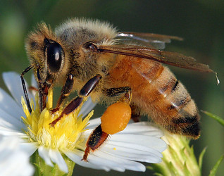
Characteristics of the host

The symbiont relationship with N. ceranae and the honey bee originated in the Asian honey bee Apis cerana, but has since then switched host to infect the European honey bee Apis mellifera. Nosema ceranae infects most of the honey bees found in Europe and North America [3].
Host-Symbiont Interaction
N. ceranae and the honeybee have a obligate parasitic relationship, meaning that the N. ceranae benefits while the honey bee is harmed throughout the process. N. ceranae forms spores which are then ingested by the honey bee through water or food. These spores then invade the gut epithelium immediately [3]. However, it is not until day six or seven that symptoms are visible such as ecoming less active, immobile, having an increase in appetite, and usually by day eight mortality has occurred [6,8]. The spores rapidly multiply in the gut and then are excreted and the spores are transferred to the other honeybees that live in the same colony through the cleaning and feeding activities [2]. Nosema ceranae decrease the ability of the honey bee to obtain nutrients from the environment which ultimately shortens their lifespan. Nosema ceranae can effect the overall colony growth and hinder winter survival [3].
Molecular Insights into the Symbiosis
There is no specific gene known to be disrupted or the cause of this infection. N. ceranae produces spores which then invade the cell, however molecular techniques such as Polymerase Chain Reaction (PCR), which amplifies a certain gene or part of a gene, allows us to see how the host cell is being effected. For example by using PCR and amplifying the 16S rRNA gene researchers are able to detect coinfection of the host by two different species [7].
Ecological and Evolutionary Aspects
This symbiont relationship between Nosema ceranae and Apis mellifera is a recent discovery, and new studies are continually done to uncover more about this parasitic relationship. Originally Nosema ceranae infected the Asian honey bee (Apis cerana), but has since then swtiched to the European honey bee (Apis mellifera). It is unknown when and how this switch occurred, and researchers are unsure if the predominant parasite of honey bees in Europe and North America still remains N. apis or if N. ceranae has taken over [3]. The biological cycle of Nosema ceranae relies on the temperature. Nosema ceranae effects more cells when kept at a constant temperature, but is a better adapter to conditions the other species such as Nosema apis. N. ceranae unlike N. apis can infect their host in all four seasons, while N. apis usually only infects their host in the milder seasons such as spring and autumn [5].
Recent Discoveries

In past studies it was thought that N. ceranae was becoming the more prevalent parasite to the Honey bee, however recent studies have shown otherwise. N. apis in a 5 year long study showed to grow in greater quantity then N. ceranae except in one colony. However it was also found that N. ceranae is a more general infection, after displaying the ability to grow in other tissues then just the gut. In this five year long study N. ceranae was observed to grow in not only the gut, but the hypopharyneal glands, salivary glands, malpighian tubes, fat bodies, and the brain. This could account for the hypothesis that N. ceranae causes more deaths in honey bee colonies then N. apis. It was also found that N. ceranae during the germination process when exposed to low temperature decreased to less the 10 percent. The remaining spores that where able to germinate were unusually short and most likely did not complete the true germination process [4].
References
[Sample reference] [[2] Seemanapalli SV, Xu Q, McShan K, Liang FT. 2010. Outer surface protein C is a dissemination-facilitating factor of Borrelia burgdorferi during mammalian infection. PLoS One 5:e15830.]
[1] http://www.diark.org/img/species_pict/Nosema_ceranae_BRL01
[2]Chen,Y., Evans,J., Murphy,C., Gutell,R., Zuker,M., Gundensen-Rindal,D and Pettis,J. 2009. Morphological, molecular, and phylogenetic characterization of Nosema ceranae, a microsporidian parasite isolated from the European honey bee, Apis mellifera. Journal of Eukaryotic Microbiology 56: 142-147.
[3]Cornman, R., Chen, Y., Schatz,M., Street,C., Zhao,Y., Desany, B., Egholm, M., Hutchison, S., Pettis, J., Lipkin and W., Eva, J. 2009. Genomic Analyses of the Microsporidian Nosema ceranae, an Emergent Pathogen of Honey Bees. PLoS Pathogens 5(6): e1000466.
[4]Gisder S., Hedtke K., Möckel N., Frielitz MC, Linde A, and Genersch E. 2010. Five-year cohort study of Nosema spp. in Germany: does climate shape virulence and assertiveness of Nosema ceranae? Applied Environmental Microbiology 76(9): 3032-3038.
[5]Hernandez, R., Meana,A., Palencia,P., Marín, P., Botías,C., Bailon, E., Barrios, L. and Higes, M., 2009. Effect of Temperature on the Biotic Potential of Honeybee Microsporidia. Applied and Enviromental Microbiology 75(8):2554.
[6]Higes, M., Garcia-Palencia, P., Martin-Hernandez, R. and Meana, A. 2006. Experimental infection of Apis mellifera honeybees with Nosema ceranae (Microsporidia). Journal of Invertbrate Pathology 94: 211-217.
[7]Higes,M., Salvador, A., Garrido-Bailón, E., Martín-Hernández,R., Meana, A. and Prieto, L. 2007. Outcome of Colonization of Nosema ceranaeby Apis mellifera. Applied and Environmental Microbiology 73(20):6331.
[8]Mayack, C. and Naug, D. 2009 Energetic stress in the honeybee Apis mellifera from Nosema ceranae infection. Journal of Invertebrate Pathology 100:185–188.
[9]http://tolweb.org/tree/ToLimages/Apis_mellifera2116051.250a.jpg
Edited by [Lacey Berry], student of Grace Lim-Fong

![[1]](http://www.kimesapiary.com/images/NosemaCeranae.jpg)