Phyllosphere: Difference between revisions
No edit summary |
No edit summary |
||
| Line 1: | Line 1: | ||
{{Uncurated}} | {{Uncurated}} | ||
= | =Introduction= | ||
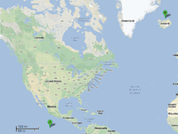
To date, the genus ''Ignicoccus'' is comprised of single cells that are irregularly shaped coccoid ranging in diameter from 1-3 µm. The Achaean genus was first isolated from [http://en.wikipedia.org/wiki/Hydrothermal_vent marine hydrothermal vents] from [http://en.wikipedia.org/wiki/Kolbeinsey Kolbeinsey Ridge] in north Iceland and also off the coast of Mexico [[#References|[1]]] (see Figure 1). They were found to have a novel cell envelope unseen before in other Achaea [[#References|[2]]], and have a very complex and poorly understood symbiotic relationship with [http://en.wikipedia.org/wiki/Nanoarchaeum_equitans ''Nanoarchaeum equitans'' ] [[#References|[3]]] [[#References|[4]]] [[#References|[5]]] [[#References|[6]]]. | |||
[[File:Ignicoccus locations.png|200px|thumb|right| Map showing the discovery locations of ''I.pacificus '' (A), and ''I.islandicus '' and '' I.hospitalis '' (B).]] | |||
= | =Current Species= | ||
There are three officially recognized ''Ignicoccus'' species: ''Ignicoccus hospitalis'' , '' Ignicoccus pacificus '' and '' Ignicoccus islandicus'' . The three species were initially identified by 16S rRNA gene analysis from the hydrothermal vent samples obtained from Kolbeinsey Ridge and the coast of Mexico[[#References|[1]]] . All three species have been characterized as hyperthermophiles that are also [http://en.wikipedia.org/wiki/Obligate_anaerobe obligate anaerobes] which explains the presence of ''Ignicoccus'' species near hydrothermal vents[[#References|[1]]] . None of the members of the ''Ignicoccus'' genus have been found to be [http://en.wikipedia.org/wiki/Pathogenic] pathogenic to humans. | |||
=Morphology= | |||
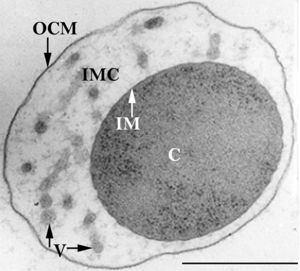
The members of the ''Ignicoccus'' genus are motile irregular coccoid cells that range in diameter from 1 to 3 µm. The motility observed is due to the presence of flagella, but unfortunately the polarity of the flagella is not yet fully elucidated. They are known to have an outer-membrane but no [http://en.wikipedia.org/wiki/S-layer S-layer]. This is a novel characteristic for these [http://en.wikipedia.org/wiki/Archaea Archaea] because''Ignicoccus'' are the only known Archaea that have been shown to possess an outer-membrane[[#References|[2]]] [[#References|[10]]] . | |||
[[File:Huber-abb2.jpg|300px|thumb|left| Ultrathin section of an ''Ignicoccus hospitalis'' cell.]] | |||
==Outer-Membrane== | |||
The outer-membrane of ''Ignicoccus'' species was found to be composed of various derivatives of the typical lipid [http://en.wikipedia.org/wiki/Archaeol archaeol], including the derivative known as [http://en.wikipedia.org/wiki/Caldarchaeol caldarchaeol] [[#References|[5]]] . The outer-membrane is dominated by a pore composed of the Imp1227 protein (''Ignicoccus'' outer membrane protein 1227). The Imp1227 protein forms a large nonamer ring with a predicted pore size of 2nm[[#References|[7]]] . | |||
= | =Metabolism= | ||
[ | ''Ignicoccus'' species are [http://en.wikipedia.org/wiki/Chemolithoautotroph chemolithoautotrophs] that use molecular hydrogen as the inorganic electron donor and elemental sulphur as the inorganic terminal electron acceptor[[#References|[1]]] . The reduction of the elemental sulphur results in the production of hydrogen sulphide gas. | ||
''Ignicoccus'' are autotrophs in that they fix their own carbon dioxide into organic molecules. The carbon dioxide fixation process they use is a novel process called [http://www.pnas.org/content/105/22/7851.full a dicarboxylate/4-hydroxybutyrate autotrophic carbon assimilation cycle] that involves 14 different enzymes[[#References|[8]]] . | |||
Members of the ''Ignicoccus'' genus are able to use ammonium as a nitrogen source. | |||
==Growth Conditions== | |||
Because members of the ''Ignicoccus'' genus are [http://en.wikipedia.org/wiki/Hyperthermophile hyperthermophiles] and obligate anaerobes, it is not surprising that their growth conditions are very complex. They are grown in a liquid medium known as ½ SME ''Ignicoccus'' which is a solution of synthetic sea water which is then made anaerobic. | |||
Grown in this media at their optimal growth temperature of 90C, the members of the ''Ignicoccus'' genus typically reach a cell density of ~4x10⁷cells/mL[[#References|[1]]] . | |||
The addition of [http://en.wikipedia.org/wiki/Yeast_extract yeast extract] to the ½ SME media has been shown to stimulate the growth and increase maximum cell density achieved. The mechanism by which this is achieved is not known[[#References|[1]]] . | |||
=Symbiosis= | |||
''Ignicoccus hospitalis'' is the only member of the genus ''Ignicoccus'' that has been shown to have an extensive [http://en.wikipedia.org/wiki/Symbiosis symbiotic relationship] with another organism. | |||
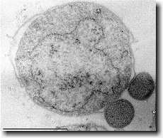
''Ignicoccus hospitalis'' has been shown to engage in symbiosis with ''Nanoarchaeum equitans'' . ''Nanoarchaeum equitans'' is a very small coccoid species with a cell diameter of 0.4 µm[[#References|[9]]] . Genome analysis has provided much of the known information about this species. | |||
To further complicate the symbiotic relationship between both species, it’s been observed that the presence of ''Nanoarchaeum equitans'' on the surface of ''Ignicoccus hospitalis'' somehow inhibits the cell replication of ''Ignicoccus hospitalis'' . How or why this occurs has not yet been elucidated[[#References|[3]]] . | |||
[[File:Urzwerg.jpg|300px|thumb|right| ''Ignicoccus hospitalis'' with two attached ''Nanoarchaeum equitans'' cells.]] | |||
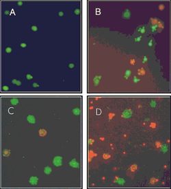
[[File:IhNeRelationship2 jpeg.jpg|250px|thumb|left| Epifluoroscence micrographs of an ''Ignicoccus hospitalis''and ''Nanoarchaeum equitans'' coculture stained with BacLight at various time points. Living cells stain green while dead cells stain red. (A) Exponential growth phase 3.25 hours after inoculation. (B) Transition into the stationary phase 7.5 hours after inoculation. (C) Stationary phase 10 hours after inoculation. (D) Stationary phase 23 hours after inoculation.]] | |||
==''Nanoarchaeum equitans''== | |||
''Nanoarchaeum equitans'' has the smallest non-viral genome ever sequenced at 491kb[[#References|[9]]] . Analysis of the genome sequence indicates that 95% of the predicted proteins and stable RNA molecules are somehow involved in repair and replication of the cell and its genome[[#References|[3]]] . | |||
Analysis of the genome also showed that ''Nanoarchaeum equitans'' lacks nearly all genes known to be required in amino acid, nucleotide, cofactor and lipid metabolism. This is partially supported by the evidence that ''Nanoarchaeum equitans'' has been shown to derive its cell membrane from its host ''Ignicoccus hospitalis'' cell membrane. The direct contact observed between ''Nanoarchaeum equitans'' and ''Ignicoccus hospitalis'' is hypothesized to form a pore between the two organisms in order to exchange metabolites or substrates (likely from ''Ignicoccus hospitalis'' towards ''Nanoarchaeum equitans'' due to the parasitic relationship). The exchange of periplasmic vesicles is not thought to be involved in metabolite or substrate exchange despite the presence of vesicles in the periplasm of ''Ignicoccus hospitalis'' . | |||
These analyses of the ''Nanoarchaeum equitans'' genome support the fact of the extensive symbiotic relationship between ''Nanoarchaeum equitans'' and ''Ignicoccus hospitalis''. However, it has not yet been proven that it is a strictly parasitic relationship and further research may prove that there is a commensal relationship between the two species. | |||
=References= | =References= | ||
(1) | (1) Burggraf S., Huber H., Mayer T., Rachel R., Stetter K.O. and Wyschkony I. ” Ignicoccus gen. nov., a novel genus of hyperthermophilic, chemolithoautotrophic Archaea, represented by two new species, Ignicoccus islandicus sp. nov. and Ignicoccus pacificus sp. nov.” International Journal of Systematic and Evolutionary Microbiology, 2000, Volume 50. | ||
(2) Naether D.J. and Rachel R. “The outer membrane of the hyperthermophilic archaeon Ignicoccus: dynamics, ultrastructure and composition.” Biochemical Society Transactions, 2004, Volume 32, part 2. | |||
(3) Giannone R.J., Heimerl T., Hettich R.L., Huber H., Karpinets T., Keller M., Kueper U., Podar M. and Rachel R. “Proteomic Characterization of Cellular and Molecular Processes that Enable the Nanoarchaeum equitans- Ignicoccus hospitalis Relationship.” PLoS ONE, 2011, Volume 6, Issue 8. | |||
(4) Eisenreich W., Gallenberger M., Huber H., Jahn U., Junglas B., Paper W., Rachel R. and Stetter K.O. “Nanoarchaeum equitans and Ignicoccus hospitalis: New Insights into a Unique, Intimate Association of Two Archaea.” Journal of Bacteriology, 2008, DOI: 10.1128/JB.01731-07. | |||
( | (5) Grosjean E., Huber H., Jahn U., Sturt H, and Summons R. “Composition of the lipids of Nanoarchaeum equitans and their origin from its host Ignicoccus sp. strain KIN4/I.” Arch Microbiol, 2004, DOI: 10.1007/s00203-004-0725-x. | ||
( | (6) Briegel A., Burghardt T., Huber H., Junglas B., Rachel R., Walther P. and Wirth R. “Ignicoccus hospitalis and Nanoarchaeum equitans: ultrastructure, cell–cell interaction, and 3D reconstruction from serial sections of freeze-substituted cells and by electron cryotomography.” Arch Microbiol, 2008, DOI 10.1007/s00203-008-0402-6. | ||
( | (7) Burghardt T., Huber H., Junglas B., Naether D.J. and Rachel R. “The dominating outer membrane protein of the hyperthermophilic Archaeum Ignicoccus hospitalis: a novel pore-forming complex.” Molecular Microbiology, 2007, Volume 63. | ||
( | (8) Berg I.A., Eisenreich W., Eylert E., Fuchs G., Gallenberger M., Huber H.,Jahn U. and Kockelkorn D. “A dicarboxylate/4-hydroxybutyrate autotrophic carbon assimilation cycle in the hyperthermophilic Archaeum Ignicoccus hospitalis.” PNAS, 2008, Volume 105, issue 22. | ||
( | (9) Brochier C., Gribaldo S., Zivanovic Y., Confalonieri F. and Forterre P. “Nanoarchaea: representatives of a novel archaeal phylum or a fast-evolving euryarchaeal lineage related to Thermococcales?” Genome Biology 2005, DOI:10.1186/gb-2005-6-5-r42. | ||
( | (10) Huber H., Rachel R., Riehl S. and Wyschkony I. “The ultrastructure of Ignicoccus: Evidence for a novel outer membrane and for intracellular vesicle budding in an archaeon.” Archaea, 2002, Volume 1. | ||
Revision as of 03:32, 14 December 2012
Introduction
To date, the genus Ignicoccus is comprised of single cells that are irregularly shaped coccoid ranging in diameter from 1-3 µm. The Achaean genus was first isolated from marine hydrothermal vents from Kolbeinsey Ridge in north Iceland and also off the coast of Mexico [1] (see Figure 1). They were found to have a novel cell envelope unseen before in other Achaea [2], and have a very complex and poorly understood symbiotic relationship with Nanoarchaeum equitans [3] [4] [5] [6].
Current Species
There are three officially recognized Ignicoccus species: Ignicoccus hospitalis , Ignicoccus pacificus and Ignicoccus islandicus . The three species were initially identified by 16S rRNA gene analysis from the hydrothermal vent samples obtained from Kolbeinsey Ridge and the coast of Mexico[1] . All three species have been characterized as hyperthermophiles that are also obligate anaerobes which explains the presence of Ignicoccus species near hydrothermal vents[1] . None of the members of the Ignicoccus genus have been found to be [1] pathogenic to humans.
Morphology
The members of the Ignicoccus genus are motile irregular coccoid cells that range in diameter from 1 to 3 µm. The motility observed is due to the presence of flagella, but unfortunately the polarity of the flagella is not yet fully elucidated. They are known to have an outer-membrane but no S-layer. This is a novel characteristic for these Archaea becauseIgnicoccus are the only known Archaea that have been shown to possess an outer-membrane[2] [10] .
Outer-Membrane
The outer-membrane of Ignicoccus species was found to be composed of various derivatives of the typical lipid archaeol, including the derivative known as caldarchaeol [5] . The outer-membrane is dominated by a pore composed of the Imp1227 protein (Ignicoccus outer membrane protein 1227). The Imp1227 protein forms a large nonamer ring with a predicted pore size of 2nm[7] .
Metabolism
Ignicoccus species are chemolithoautotrophs that use molecular hydrogen as the inorganic electron donor and elemental sulphur as the inorganic terminal electron acceptor[1] . The reduction of the elemental sulphur results in the production of hydrogen sulphide gas.
Ignicoccus are autotrophs in that they fix their own carbon dioxide into organic molecules. The carbon dioxide fixation process they use is a novel process called a dicarboxylate/4-hydroxybutyrate autotrophic carbon assimilation cycle that involves 14 different enzymes[8] .
Members of the Ignicoccus genus are able to use ammonium as a nitrogen source.
Growth Conditions
Because members of the Ignicoccus genus are hyperthermophiles and obligate anaerobes, it is not surprising that their growth conditions are very complex. They are grown in a liquid medium known as ½ SME Ignicoccus which is a solution of synthetic sea water which is then made anaerobic.
Grown in this media at their optimal growth temperature of 90C, the members of the Ignicoccus genus typically reach a cell density of ~4x10⁷cells/mL[1] .
The addition of yeast extract to the ½ SME media has been shown to stimulate the growth and increase maximum cell density achieved. The mechanism by which this is achieved is not known[1] .
Symbiosis
Ignicoccus hospitalis is the only member of the genus Ignicoccus that has been shown to have an extensive symbiotic relationship with another organism.
Ignicoccus hospitalis has been shown to engage in symbiosis with Nanoarchaeum equitans . Nanoarchaeum equitans is a very small coccoid species with a cell diameter of 0.4 µm[9] . Genome analysis has provided much of the known information about this species.
To further complicate the symbiotic relationship between both species, it’s been observed that the presence of Nanoarchaeum equitans on the surface of Ignicoccus hospitalis somehow inhibits the cell replication of Ignicoccus hospitalis . How or why this occurs has not yet been elucidated[3] .

Nanoarchaeum equitans
Nanoarchaeum equitans has the smallest non-viral genome ever sequenced at 491kb[9] . Analysis of the genome sequence indicates that 95% of the predicted proteins and stable RNA molecules are somehow involved in repair and replication of the cell and its genome[3] .
Analysis of the genome also showed that Nanoarchaeum equitans lacks nearly all genes known to be required in amino acid, nucleotide, cofactor and lipid metabolism. This is partially supported by the evidence that Nanoarchaeum equitans has been shown to derive its cell membrane from its host Ignicoccus hospitalis cell membrane. The direct contact observed between Nanoarchaeum equitans and Ignicoccus hospitalis is hypothesized to form a pore between the two organisms in order to exchange metabolites or substrates (likely from Ignicoccus hospitalis towards Nanoarchaeum equitans due to the parasitic relationship). The exchange of periplasmic vesicles is not thought to be involved in metabolite or substrate exchange despite the presence of vesicles in the periplasm of Ignicoccus hospitalis .
These analyses of the Nanoarchaeum equitans genome support the fact of the extensive symbiotic relationship between Nanoarchaeum equitans and Ignicoccus hospitalis. However, it has not yet been proven that it is a strictly parasitic relationship and further research may prove that there is a commensal relationship between the two species.
References
(1) Burggraf S., Huber H., Mayer T., Rachel R., Stetter K.O. and Wyschkony I. ” Ignicoccus gen. nov., a novel genus of hyperthermophilic, chemolithoautotrophic Archaea, represented by two new species, Ignicoccus islandicus sp. nov. and Ignicoccus pacificus sp. nov.” International Journal of Systematic and Evolutionary Microbiology, 2000, Volume 50.
(2) Naether D.J. and Rachel R. “The outer membrane of the hyperthermophilic archaeon Ignicoccus: dynamics, ultrastructure and composition.” Biochemical Society Transactions, 2004, Volume 32, part 2.
(3) Giannone R.J., Heimerl T., Hettich R.L., Huber H., Karpinets T., Keller M., Kueper U., Podar M. and Rachel R. “Proteomic Characterization of Cellular and Molecular Processes that Enable the Nanoarchaeum equitans- Ignicoccus hospitalis Relationship.” PLoS ONE, 2011, Volume 6, Issue 8.
(4) Eisenreich W., Gallenberger M., Huber H., Jahn U., Junglas B., Paper W., Rachel R. and Stetter K.O. “Nanoarchaeum equitans and Ignicoccus hospitalis: New Insights into a Unique, Intimate Association of Two Archaea.” Journal of Bacteriology, 2008, DOI: 10.1128/JB.01731-07.
(5) Grosjean E., Huber H., Jahn U., Sturt H, and Summons R. “Composition of the lipids of Nanoarchaeum equitans and their origin from its host Ignicoccus sp. strain KIN4/I.” Arch Microbiol, 2004, DOI: 10.1007/s00203-004-0725-x.
(6) Briegel A., Burghardt T., Huber H., Junglas B., Rachel R., Walther P. and Wirth R. “Ignicoccus hospitalis and Nanoarchaeum equitans: ultrastructure, cell–cell interaction, and 3D reconstruction from serial sections of freeze-substituted cells and by electron cryotomography.” Arch Microbiol, 2008, DOI 10.1007/s00203-008-0402-6.
(7) Burghardt T., Huber H., Junglas B., Naether D.J. and Rachel R. “The dominating outer membrane protein of the hyperthermophilic Archaeum Ignicoccus hospitalis: a novel pore-forming complex.” Molecular Microbiology, 2007, Volume 63.
(8) Berg I.A., Eisenreich W., Eylert E., Fuchs G., Gallenberger M., Huber H.,Jahn U. and Kockelkorn D. “A dicarboxylate/4-hydroxybutyrate autotrophic carbon assimilation cycle in the hyperthermophilic Archaeum Ignicoccus hospitalis.” PNAS, 2008, Volume 105, issue 22.
(9) Brochier C., Gribaldo S., Zivanovic Y., Confalonieri F. and Forterre P. “Nanoarchaea: representatives of a novel archaeal phylum or a fast-evolving euryarchaeal lineage related to Thermococcales?” Genome Biology 2005, DOI:10.1186/gb-2005-6-5-r42.
(10) Huber H., Rachel R., Riehl S. and Wyschkony I. “The ultrastructure of Ignicoccus: Evidence for a novel outer membrane and for intracellular vesicle budding in an archaeon.” Archaea, 2002, Volume 1.


