File list
From MicrobeWiki, the student-edited microbiology resource
This special page shows all uploaded files.
| Date | Name | Thumbnail | Size | User | Description | Versions |
|---|---|---|---|---|---|---|
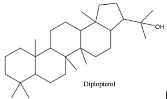
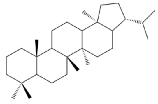
| 18:43, 24 April 2018 | Diplopterol.png (file) |  |
39 KB | Li1 | 1 | |
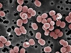
| 17:00, 24 April 2018 | Enterococcus-faecium-250x250.jpg (file) |  |
14 KB | Millermr | SEM of E. faecium | 1 |
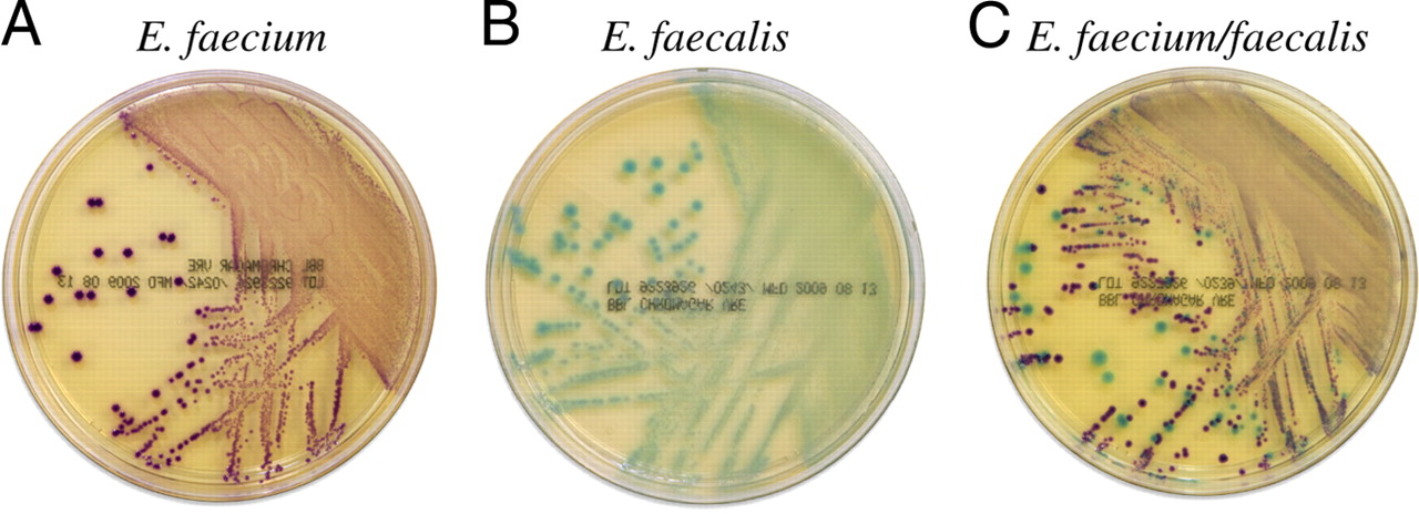
| 16:59, 24 April 2018 | E faecium vs E faecalis.jpg (file) |  |
132 KB | Millermr | E. faecium and E. faecalis on BBL CHROMagar VanRE. | 1 |
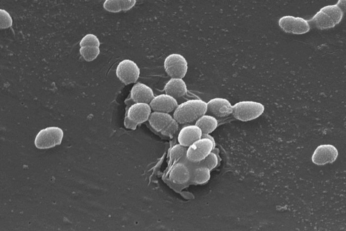
| 16:58, 24 April 2018 | Enterococcus-faecalis.png (file) |  |
527 KB | Millermr | E. faecalis SEM image | 1 |
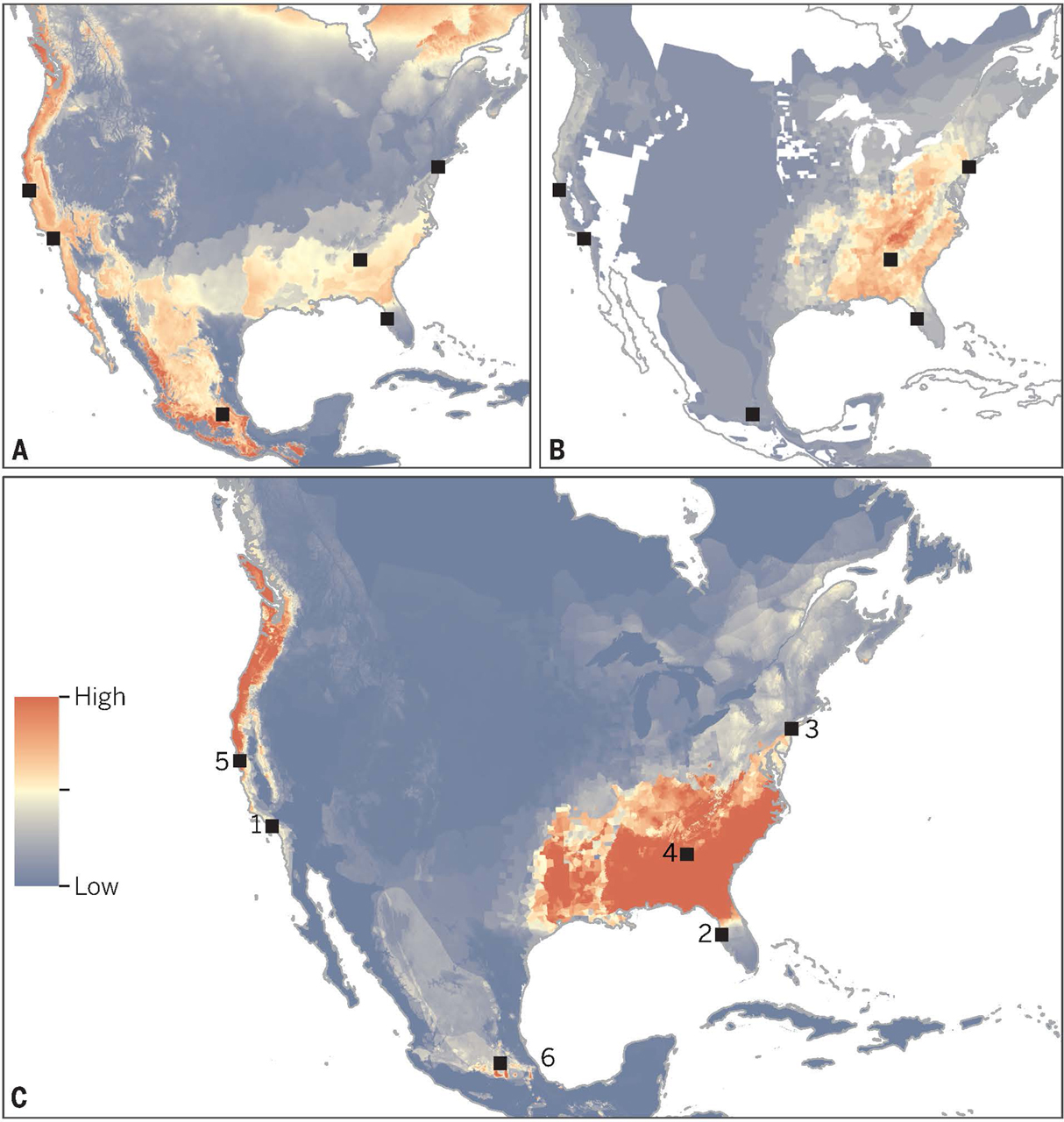
| 14:05, 24 April 2018 | Yap Map of Vulnerability.jpg (file) |  |
625 KB | Ellsworth1 | 1 | |
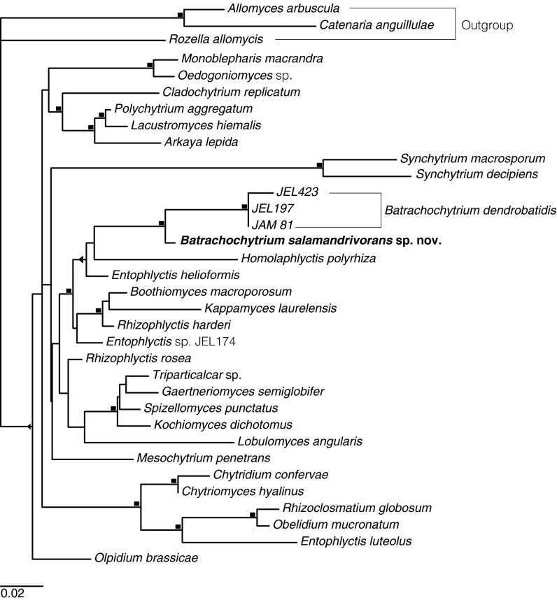
| 14:00, 24 April 2018 | Martel Etymology.jpg (file) |  |
108 KB | Ellsworth1 | 1 | |
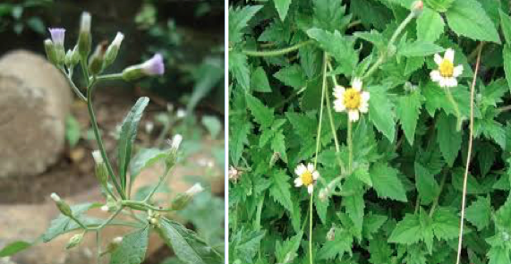
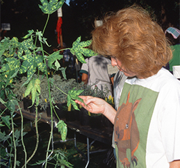
| 13:58, 24 April 2018 | Dengueplants.png (file) |  |
362 KB | Vitalen | 1 | |
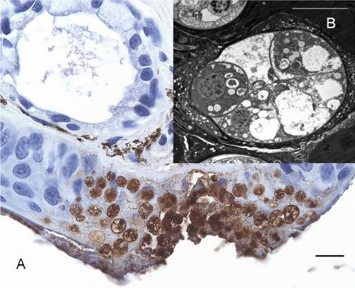
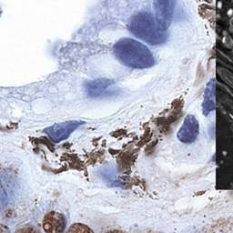
| 13:56, 24 April 2018 | Martel Skin lesions.jpg (file) |  |
44 KB | Ellsworth1 | 1 | |
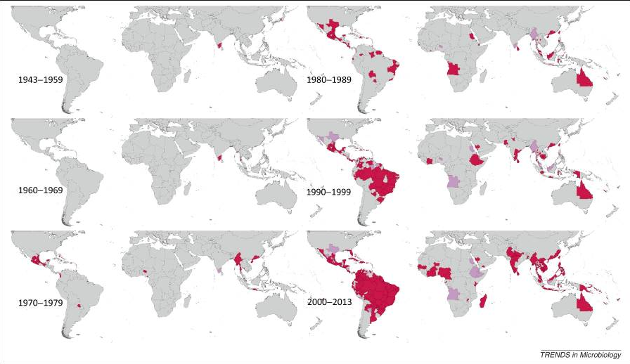
| 13:50, 24 April 2018 | Denguemap.png (file) |  |
392 KB | Vitalen | 1 | |
| 13:50, 24 April 2018 | Martel Skin leisons.jpg (file) |  |
13 KB | Ellsworth1 | 1 | |
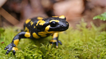
| 13:40, 24 April 2018 | Fire Salamander Google Images.jpg (file) |  |
28 KB | Ellsworth1 | 1 | |
| 03:41, 24 April 2018 | D-WO-ABMV-FO.003b.jpg (file) |  |
113 KB | Manleyd | A plant infected with AbMV for commercial sale being inspected by customer. | 1 |
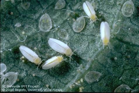
| 03:40, 24 April 2018 | I-HO-BTAB-AD.006.jpg (file) |  |
34 KB | Manleyd | Sweet Potato Whitefly | 1 |
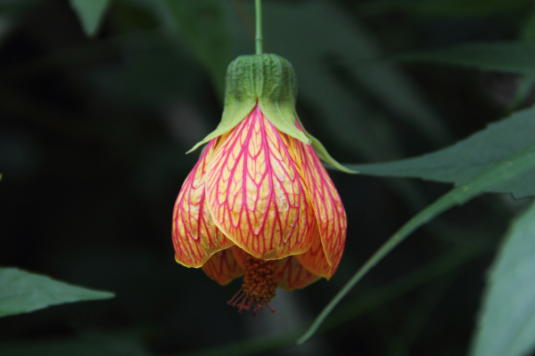
| 03:38, 24 April 2018 | Abutilon striatum1200.jpg (file) |  |
156 KB | Manleyd | Abutilon striatum | 1 |
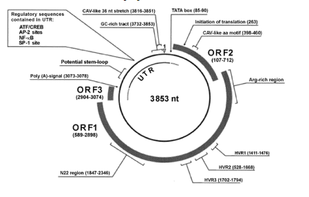
| 03:36, 24 April 2018 | CaMV genome.jpg (file) |  |
62 KB | Brennana | CaMV Genome | 1 |
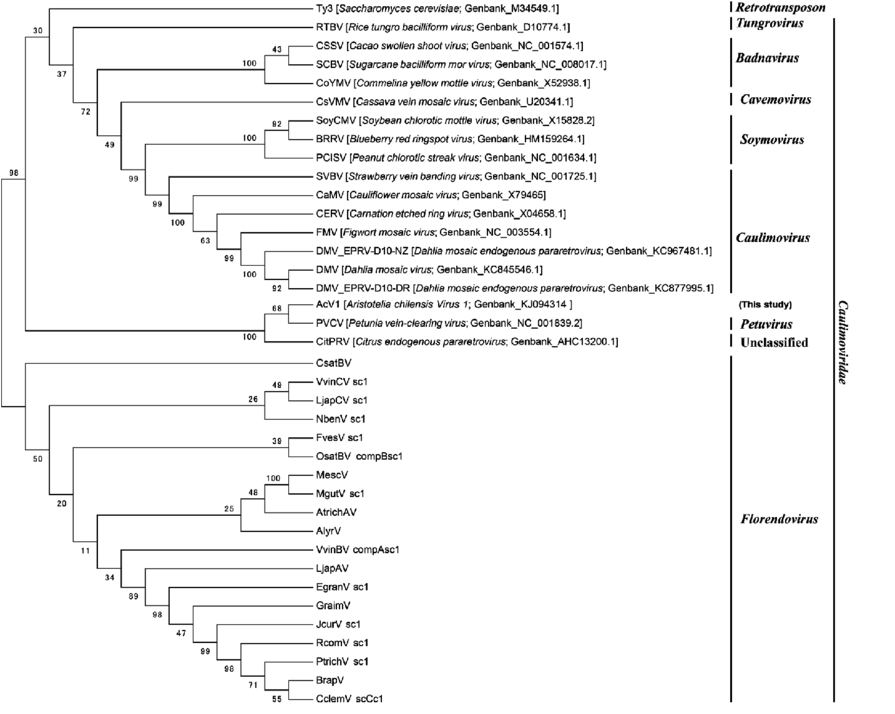
| 03:32, 24 April 2018 | Cauliflower Mosaic Virus Phylogeny.png (file) |  |
133 KB | Brennana | CaMV Phylogeny | 1 |
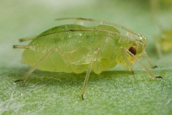
| 03:28, 24 April 2018 | Myszus-persicae.jpg (file) |  |
80 KB | Brennana | Myszus persicae | 1 |
| 03:21, 24 April 2018 | Skeleton.png (file) |  |
38 KB | Li1 | 1 | |
| 03:17, 24 April 2018 | Bile salts.png (file) |  |
503 KB | Li1 | 1 | |
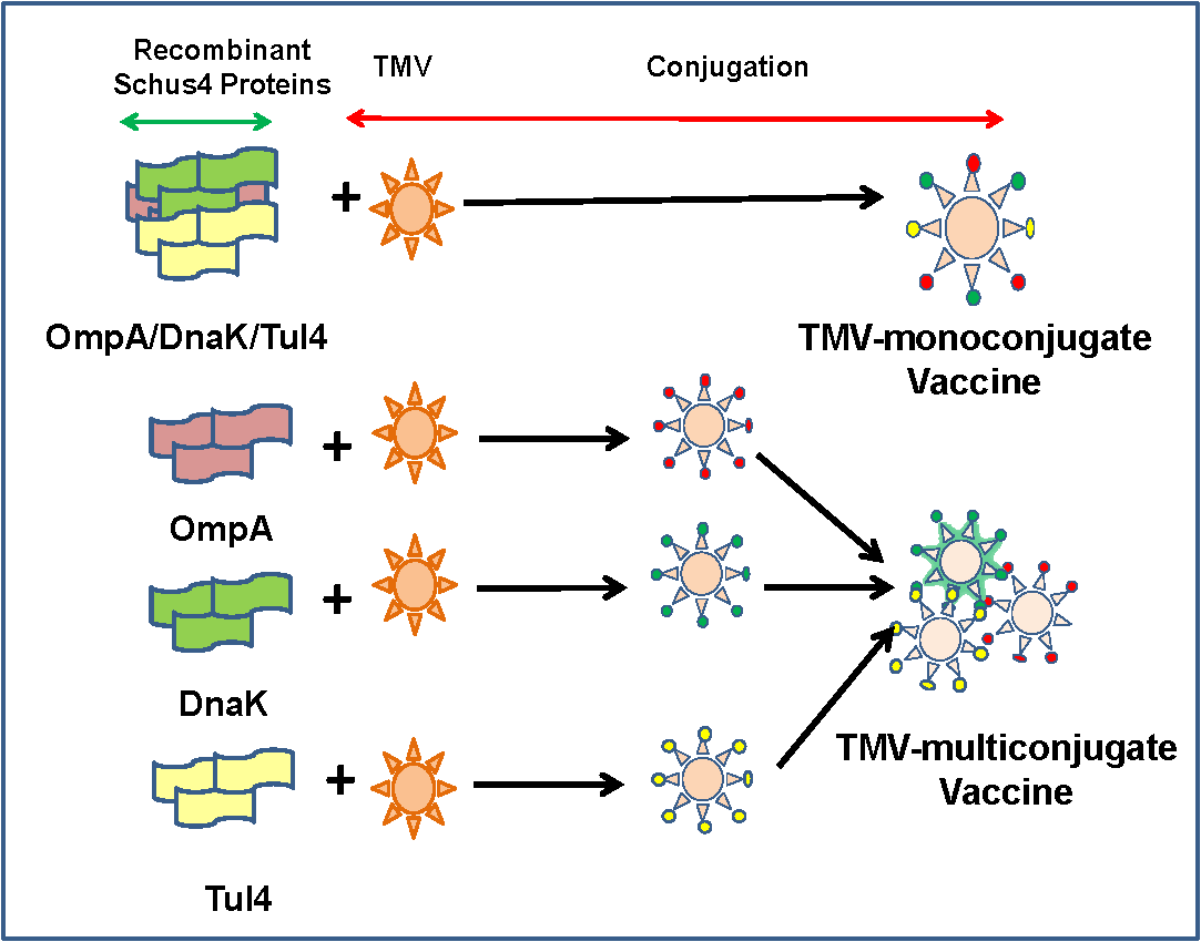
| 02:19, 24 April 2018 | VaccineTMV.PNG (file) |  |
32 KB | Itschnerms | 1 | |
| 02:10, 24 April 2018 | TMVmelanoma.jpg (file) |  |
391 KB | Itschnerms | 1 | |
| 01:51, 24 April 2018 | SEMelectrodesTMV.gif (file) |  |
196 KB | Itschnerms | 1 | |
| 01:43, 24 April 2018 | TMV.jpg (file) |  |
61 KB | Itschnerms | 1 | |
| 01:34, 24 April 2018 | 13337 2010 7 Fig1 HTML.jpg (file) |  |
88 KB | Tosaris1 | Dog exposed to canine parvovirus. Intravenous and subcutaneous drip sets hydrating dog. | 1 |
| 01:29, 24 April 2018 | Screen Shot 2018-04-23 at 9.28.45 PM.png (file) |  |
1.03 MB | Tosaris1 | VP2 gene location sequencing alignment of two dog communities, Parvo6 and Parvo9. Parvo6 and Parvo9 chosen to compare with reference VP2 amino acid structure. Analysis of 18 amino acids at each subtype revealed similarities between the two communities... | 1 |
| 01:26, 24 April 2018 | Screen Shot 2018-04-23 at 9.22.55 PM.png (file) |  |
88 KB | Tosaris1 | Virus isolation shown without and without (+/-) VN antibodies. Feline subjects inoculated with reference FPLV, CPV-2a and CPV-2c. Blood samples taken at the time of inoculation. | 1 |
| 01:21, 24 April 2018 | Diagrammatic-representation-of-the-intestinal-epithelia-A-transverse-section-of-the.jpg (file) | 55 KB | Tosaris1 | Drawing of microscopic epithelial cell in the gastrointestinal tract. Crypt intestinal gland involved in reproduction of new cells that migrate upward from the gland into the lumen. | 1 | |
| 00:31, 24 April 2018 | Epithelial cell migration.tif.png (file) |  |
32 KB | Tosaris1 | Microscopic drawn rendering of epithelial cell in gastrointestinal tract. Crypt intestinal gland involved in the proliferation of new cells into the lumen. | 1 |
| 19:30, 23 April 2018 | Ismej20138f9.jpg (file) |  |
88 KB | Hanson1 | 1 | |
| 19:28, 23 April 2018 | Ismej20138f3.jpg (file) | 20 KB | Hanson1 | 1 | ||
| 15:55, 23 April 2018 | Dead.jpg (file) |  |
83 KB | Kotnourj | 1 | |
| 15:49, 23 April 2018 | Penguin.jpg (file) |  |
117 KB | Kotnourj | 1 | |
| 13:05, 23 April 2018 | Manzin et al. genome stx.png (file) |  |
53 KB | Josowitzj | 1 | |
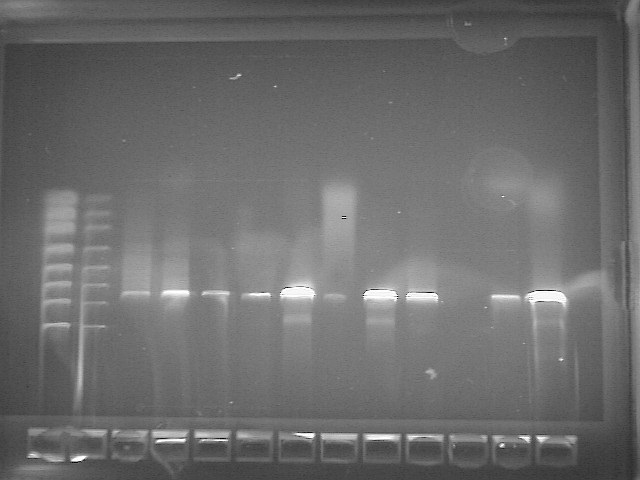
| 20:05, 22 April 2018 | PCR Bacillus Subtilus.jpg (file) |  |
67 KB | Nathan.zuck | 1 | |
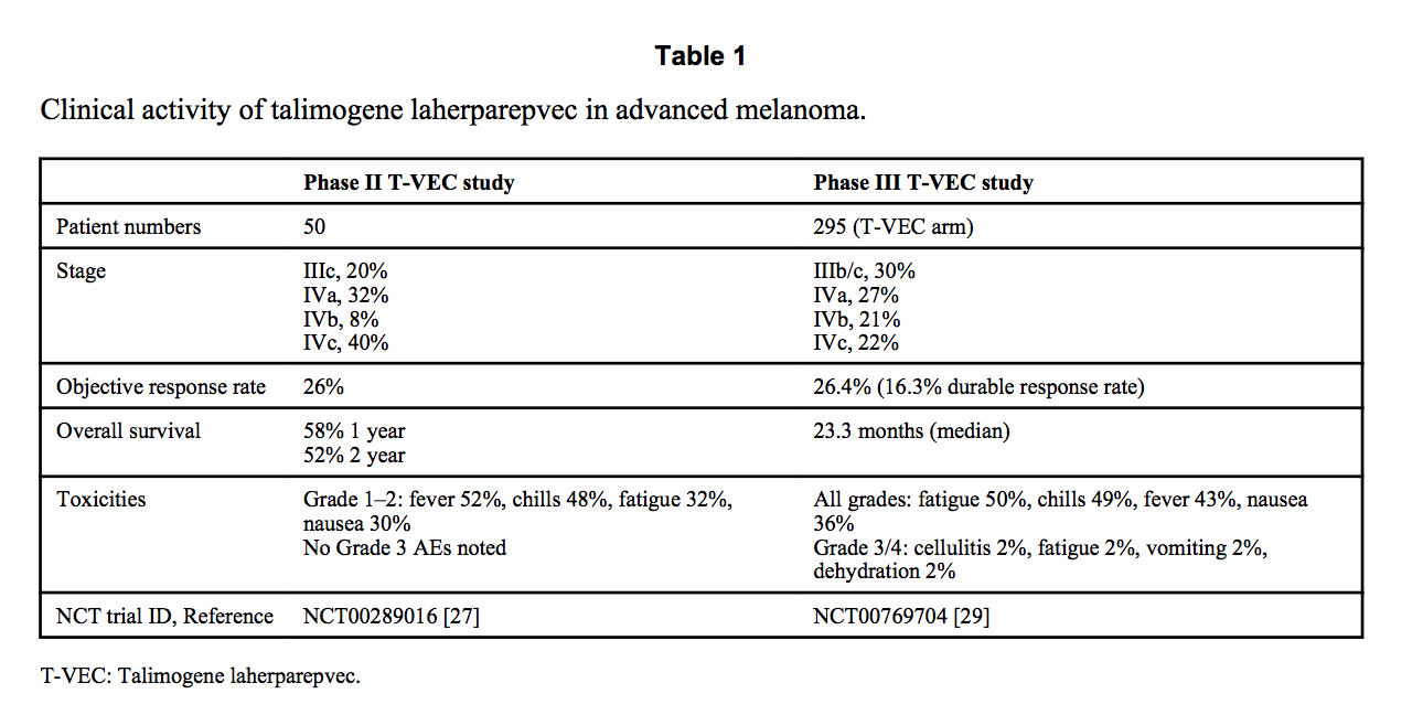
| 16:46, 22 April 2018 | Tvectable1.png (file) |  |
118 KB | Faziolia | 1 | |
| 16:40, 22 April 2018 | TVECM.png (file) |  |
516 KB | Faziolia | 1 | |
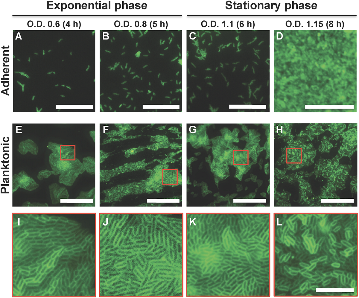
| 23:30, 19 April 2018 | Free floating p stuartii.PNG (file) |  |
1.67 MB | Labunish | [https://doi.org/10.1371/journal.pone.0174213 El Khatib M, Tran Q-T, Nasrallah C, Lopes J, Bolla J-M, Vivaudou M, et al. (2017) Providencia stuartii form biofilms and floating communities of cells that display high resistance to environmental insults.... | 1 |
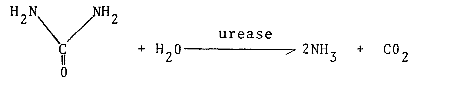
| 23:13, 19 April 2018 | Ureaserxn.png (file) | 3 KB | Labunish | from https://data.epo.org/publication-server/rest/v1.0/publication-dates/19810701/patents/EP0031051NWA1/document.html | 1 | |
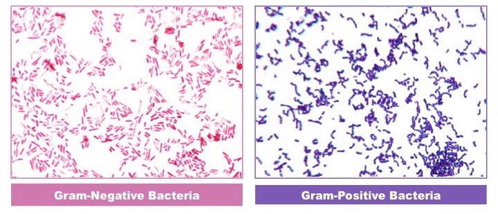
| 22:52, 19 April 2018 | Negvposbact.jpeg (file) |  |
83 KB | Labunish | Visual comparison of gram-negativE bacteria and gram-positive bacteria using the gram-staining technique (NIAID) | 1 |
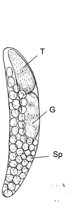
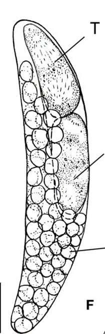
| 15:20, 19 April 2018 | Fullygrownparasite.png (file) |  |
29 KB | Corhoski | Figure 1 shows a fully grown parasite which would live inside its host. (T) points to the trophocyte, (G) points to the gonocyte, and (Sp) shows the sporocytes. (Image credited to frontiers in microbiology, https://www.ncbi.nlm.nih.gov/pmc/articles/PMC... | 2 |
| 14:53, 19 April 2018 | FullyGrown.png (file) |  |
44 KB | Corhoski | 1 | |
| 14:51, 19 April 2018 | Fully grown.png (file) |  |
44 KB | Corhoski | 1 | |
| 14:50, 19 April 2018 | Bs pic 1.jpg (file) |  |
190 KB | Ellsworth1 | 1 | |
| 01:46, 19 April 2018 | Granulosis.gif (file) |  |
126 KB | Ikundel | 1 | |
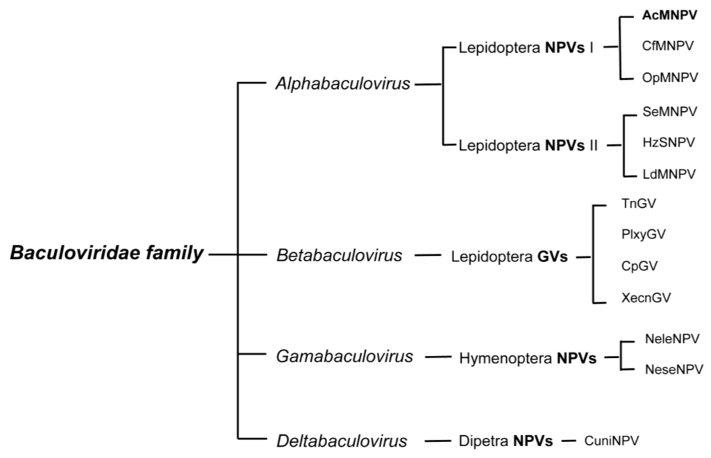
| 00:03, 19 April 2018 | Baculovirus taxonomy.png (file) |  |
51 KB | Ikundel | 1 | |
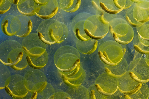
| 21:15, 18 April 2018 | Oophila2.jpg (file) |  |
136 KB | Mzaret | 1 | |
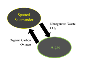
| 21:02, 18 April 2018 | Oophila1.png (file) |  |
18 KB | Mzaret | 1 | |
| 20:59, 18 April 2018 | 108ef8b8771ca41a65d7acc195ab25f9.jpg (file) |  |
281 KB | Jesszell1 | 1 | |
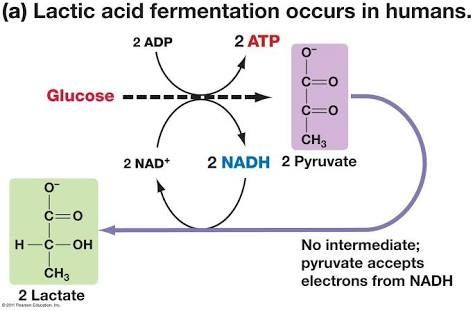
| 16:15, 18 April 2018 | Main-qimg-2d62a355a4c1fc399a411a1093c69f44-c.jpg (file) |  |
21 KB | Ppprocto | Image taken from Wewin Tjiasmanto at https://www.quora.com/What-are-the-main-steps-of-lactic-acid-fermentation | 1 |
| 15:25, 18 April 2018 | Abutilon-mosaic.jpg (file) |  |
865 KB | Slonczewski | Leaves infected with Abutilon mosaic virus Source: https://rybicki.files.wordpress.com/2012/02/abutilon-mosaic.jpg | 1 |
