|
|
| (45 intermediate revisions by 2 users not shown) |
| Line 1: |
Line 1: |
| | {{Uncurated}} |
| {{Biorealm Phylum}} | | {{Biorealm Phylum}} |
| | |
| | |
| | [[Image:vert1.gif|thumb|350px|right| Verticillium sp. well differentiated and erect conidiophores, verticillately branched over most of their length, bearing whorls of slender awl-shaped divergent phialides. Conidia are hyaline or brightly coloured, mostly one-celled, and are usually borne in slimy heads (glioconidia). |
| | [http://www.mycology.adelaide.edu.au/gallery/photos/vert1.html]]] |
| | |
|
| |
|
| <h2>Classification</h2> | | <h2>Classification</h2> |
| Line 14: |
Line 20: |
|
| |
|
| <h2>Description and Significance</h2> | | <h2>Description and Significance</h2> |
| Species within the Zygomycota classification make up only about 1% of true Fungi. There are only about 900 species. However, humans rarely encounter most species. The most familiar is the mold that affects strawberries and other fruits. This phylum encompasses at least seven orders. Zygomycota are commonly thought of as bread molds, but there are many species of fungi within this classification that form symbiotic relationships with plants or infect animal hosts. Two other common names for Zygomycota are pin molds and sugar molds. The term "pin mold" refers to the appearance of certain species, while "sugar molds" refers to the sugar-rich fruit that is often affected by zygomycota. They are thought to be the most primitive terrestrial fungi. It is believed that Zygomycota emerged between 600 and 1,400 million years ago. It is suggested that Zygomycota are either para- or polyphletic, but this has yet to be determine. They share many characteristics with flagellated fungi, and therefore were once thought to be related to acquatic fungi. However, differences in cell-wall structure and a lack of flagellated spores or gamets indicate that there is no relation.
| | [[Image:3122_5.jpg|thumb|250px|right|One-sided wilt and death of potato leaves caused by Verticillium spp''. by: [http://ohioline.osu.edu/hyg-fact/3000/3122.html]]] |
| | [[Image:3122_4.jpg|thumb|250px|left|Typical V-shaped lesions on tomato leaves associated with Verticillium wilt''. by: [http://ohioline.osu.edu/hyg-fact/3000/3122.html]]] |
| | "Verticillium is a filamentous fungus that inhabits decaying vegetation and soil. Some Verticillium species may be pathogenic to arthropods, plants, and other fungi. It is commonly considered as a contaminant. Verticillium may very rarely cause human disease."[1] |
| | |
| | "Verticillium is also a genus of fungi of [[Ascomycota]]. This genus has three ecologically based group: mycopathogens, entomopathogens, and plant pathogens and related saprophytes."[7] The Verticillium belongs to the plant pathogens group. |
| | |
| | Verticillim dehliae and Verticillim albo-atrum cause wilt diseases, which refers to loss of rigidity of non-woody parts of plants, in plants such as "cotton, potatos, peppers, eggplants, tomatoes, and ornamental woody plants"[7]. Because Verticillium species are soil-borne and can attack both herbaceous and woody plants, the Verticillium wilt is difficult to control.[4] |
| | |
| | [[Image:VerticilliumWilt.jpg|thumb|250px|right|Symptoms of Verticillium wilt on maple: the left half of the tree appears diseased, while the right half appears healthy. |
| | (Photo by R. J. Stipes). by: [http://www.ext.vt.edu/pubs/plantdiseasefs/450-619/450-619.html]]] |
| | |
| | <h2>Microscopic Features</h2> |
| | |
| | "Septate hyaline hyphae, conidiophores, phialides, and conidia are observed. Conidiophores are hyaline, simple or branched. The branching of the conidiophores occurs in whorls at several levels. Conidiophores bear the phialides. Phialides are very long and are also arranged in verticils (whorls) around the conidiophore. Verticils may be disrupted in slide culture. The apices of the phialides are pointed. Conidia (2-13µm in length) are hyaline or brightly colored, one-celled, and oval to pyriform in shape. They are solitary or form clusters in sticky heads at the tips of the phialides." [1][3][6] |
| | |
| | <h2>Pathogenicity and Clinical Significance</h2> |
| | "Verticillium has been reported as a possible cause of keratitis in humans. "[1][2][3] |
| | |
| | <h2>References</h2> |
| | 1 http://www.doctorfungus.org/thefungi/verticillium.htm |
|
| |
|
| <h2>Genome Structure</h2>
| | 2 Larone, D. H. 1995. Medically Important Fungi - A Guide to Identification, 3rd ed. ASM Press, Washington, D.C. |
| Zygomycota is a classification that encompasses many different species with very different genome structures.
| |
|
| |
|
| <h2>Cell Structure and Metabolism</h2>
| | 3 Sutton, D. A., A. W. Fothergill, and M. G. Rinaldi (ed.). 1998. Guide to Clinically Significant Fungi, 1st ed. Williams & Wilkins, Baltimore. |
| [[image:Syncep1.jpg|frame|left|Syncephalastrum racemosum. [http://www.botany.hawaii.edu/faculty/wong/Bot201/Myxomycota/Introduction.htm Introduction to the Fungi by George Wong]]]
| |
| <br>
| |
| <br>
| |
| Cell walls are composed of chitin-chitosan. The mature zygospore has thick walls. Zygomycota also have coenocytic mycelium.
| |
|
| |
|
| | 4 http://www.ext.vt.edu/pubs/plantdiseasefs/450-619/450-619.html |
|
| |
|
| They normally grow as mycellia or as filaments of long cells. Hyphae typically lack cross walls or septa, and therefore are coeonocytic. It is believed that zygomycota have zygotic or haplontic life cycles. Zygomycota are able to reproduce both sexually and asexually. In fact, sexual reproduction via zygospores following gametangial fusion is a definitive characteristic of Zygomycota. Sexual reproduction is haploid-dominant, while asexual reproduction
| | 5 Larone, D. H. 1995. Medically Important Fungi - A Guide to Identification, 3rd ed. ASM Press, Washington, D.C. |
| makes use of aplanospores. In Zygomycota, sexual reproduction is the fusion of undifferentiated isogametangia or anisogametangia. Mating strains (progametangia) grow toward each other and induce
| |
| the hormone trisporic acid to intiate sexual development. This fusion produces a zygote, which develops into a zygospore. The way Zygomycota reproduce asexually is also distinctive. With
| |
| asexual reproduction, asexual spores called sporangiospores are produced in sporangia. Typically, three sporangia are produced, but there are variations within asexual reproduction. Nuclei
| |
| move to the ends of the progametangia to form septa. Plasmogamy and karyogamy follow the fusion of the progametangia. Sexual reproduction ends with the formation of the zygospore. Because
| |
| of the Zgomycota life cycle, only the diploid phase takes place in the zygospore. Zygospores must undergo a dormant period before beginning the reproductive cycle again.
| |
| <br style="clear:both" />
| |
| <h2>Ecology</h2>
| |
| Zygomycota are arguably the most ecologically diverse group of fungi. Zygomycota are terrestrial organisms. They live close to plants, usually in soil and on decaying plant matter. Because they decompose soil, plant matter, and dung, they have a major role in the carbon cycle. Zygomycota are also pathogens for animals, amebas, plants, and other fungi. They form mutualistic symbiotic relationships with plants. In addition, they form commensalistic relationships with arthropods, inhabiting the gut of the organism and feeding on unused nutrients. However, Zygomycota can also be found in acquatic ecosystems.
| |
| While Zygomycota are largely known to humans for the negative economic impact they have on fruit, they also have some practical use. For example, certain species are used in Asian food fermentations. In addition, people have used their pathogenic powers to control insect pests. Although these are largely considered terrestrial organisms, certain species of Zygomycota also form relationships with animals. Zygomycetes are known to cause serious infections, articularly for diabetics and immunocompromised individuals. These infections can also occur as a result of major burns or other tramatic injury. One such disease is zygomycosis. This is a rare fungal disease that occurs in humans, and can even affect the fetus. It is potentially lethal. Diven et. al. (2004) note three clinical forms: cellulitis, disseminated, and gastrointestinal. The symptoms of the gastrointestinal form mirror those of another disease, necrotizing enterocolitis (NEC). This often makes diagnosis of the disease difficult. Thammayya (2005) wrote case study on a form of the diesease that is an upper respiratory tract infection with a wide variety of symptoms. Some of these include epistaxis, intranasal tumor, and nasal obstruction. Thammayya's case study noted that it was the first report on this disease due to a species from North-eastern India. One research focus regarding the infections species is on how to control and treat them. The work of -Lamaignaire et. al. (2005) studied the affects of polymorphonuclear leukocytes (PMNLs) on certain species of zygomycetes, and compared these results to those of interferon (IFN)- gamma and anulocyte-macrophage colony-stimulating factor (GM-CSF). It was found that while the PMNLs did have ome effect, the IFN-gammas and GM-CSFs were more affective in combatting the invasive organisms.
| |
|
| |
|
| Zygomycota are not just restricted to the biological world. The modern dance company Pilobolus, founded in 1971, took its name from the fungus.
| | 6 St-Germain, G., and R. Summerbell. 1996. Identifying Filamentous Fungi - A Clinical Laboratory Handbook, 1st ed. Star Publishing Company, Belmont, California. |
|
| |
|
| [[image:31-07-ZygomyceteLifeCyc-L.jpg|frame|center|Zygomycota life cycle. [http://www.anselm.edu/homepage/jpitocch/genbios/surveybi04.html Survey of representatives of the major Kingdoms by Jay Pitocchelli.]]]
| | 7 http://en.wikipedia.org/wiki/Verticillium |
| [[image:Splash.jpg|frame|center|[http://www.pilobolus.com/ Members of Pilobolus. Photo taken by John Kane.]]]
| |
| <h2>References. Updated May 19, 2005</h2>
| |
A Microbial Biorealm page on the phylum Verticillium
Verticillium sp. well differentiated and erect conidiophores, verticillately branched over most of their length, bearing whorls of slender awl-shaped divergent phialides. Conidia are hyaline or brightly coloured, mostly one-celled, and are usually borne in slimy heads (glioconidia).
[1]
Classification
Higher order taxa
Division Eucaryota,
Kingdom Fungi,
Phylum Ascomycota,
Class Incertae sedis
Species
Verticillium dahliae
Verticillium lecanii
Verticillium albo-atrum
Description and Significance
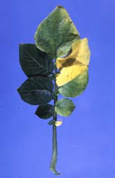
One-sided wilt and death of potato leaves caused by Verticillium spp
. by: [2]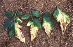
Typical V-shaped lesions on tomato leaves associated with Verticillium wilt
. by: [3]"Verticillium is a filamentous fungus that inhabits decaying vegetation and soil. Some Verticillium species may be pathogenic to arthropods, plants, and other fungi. It is commonly considered as a contaminant. Verticillium may very rarely cause human disease."[1]
"Verticillium is also a genus of fungi of Ascomycota. This genus has three ecologically based group: mycopathogens, entomopathogens, and plant pathogens and related saprophytes."[7] The Verticillium belongs to the plant pathogens group.
Verticillim dehliae and Verticillim albo-atrum cause wilt diseases, which refers to loss of rigidity of non-woody parts of plants, in plants such as "cotton, potatos, peppers, eggplants, tomatoes, and ornamental woody plants"[7]. Because Verticillium species are soil-borne and can attack both herbaceous and woody plants, the Verticillium wilt is difficult to control.[4]
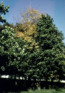
Symptoms of Verticillium wilt on maple: the left half of the tree appears diseased, while the right half appears healthy. (Photo by R. J. Stipes). by:
[4]Microscopic Features
"Septate hyaline hyphae, conidiophores, phialides, and conidia are observed. Conidiophores are hyaline, simple or branched. The branching of the conidiophores occurs in whorls at several levels. Conidiophores bear the phialides. Phialides are very long and are also arranged in verticils (whorls) around the conidiophore. Verticils may be disrupted in slide culture. The apices of the phialides are pointed. Conidia (2-13µm in length) are hyaline or brightly colored, one-celled, and oval to pyriform in shape. They are solitary or form clusters in sticky heads at the tips of the phialides." [1][3][6]
Pathogenicity and Clinical Significance
"Verticillium has been reported as a possible cause of keratitis in humans. "[1][2][3]
References
1 http://www.doctorfungus.org/thefungi/verticillium.htm
2 Larone, D. H. 1995. Medically Important Fungi - A Guide to Identification, 3rd ed. ASM Press, Washington, D.C.
3 Sutton, D. A., A. W. Fothergill, and M. G. Rinaldi (ed.). 1998. Guide to Clinically Significant Fungi, 1st ed. Williams & Wilkins, Baltimore.
4 http://www.ext.vt.edu/pubs/plantdiseasefs/450-619/450-619.html
5 Larone, D. H. 1995. Medically Important Fungi - A Guide to Identification, 3rd ed. ASM Press, Washington, D.C.
6 St-Germain, G., and R. Summerbell. 1996. Identifying Filamentous Fungi - A Clinical Laboratory Handbook, 1st ed. Star Publishing Company, Belmont, California.
7 http://en.wikipedia.org/wiki/Verticillium


