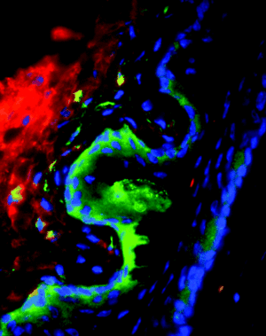
Ectromelia virus
Characteristics of Ectromelia virus

Ectromelia virus (ECTV) is a zoonotic viral disease. It belongs to the Poxviridae family of the genus Orthopoxvirus and is among the species of Vaccinia virus. Virions are oval or brick-shaped with a dimension of approximately 175 X 290 nm. It has a linear, double-stranded DNA genome that is 209,771 bp, surronded by a layer of lipids. [1.,2.,3.,4.]. This host-specialized virus infects with high efficiency, with the ability to spread systematically within its host and be effectively transmitted to others [1.,2.,5.,6.].
Characteristics of the host

The host of ECTV is Mus musculus, better known as the mouse [3.]. While all mice are susceptible to the infection, clinical disease and mortality of mice is dependent upon virus and mouse strain. Mice strains highly susceptible to ECTV include A, CBA, C3H, DBA/2, and BALB/c while those that appear to be most resistant to infection include C57BL/6 and C57BL/10. The infection is not commonly seen among commercial colonies of mice but rather in research laboratories that exchange mouse tissues, live mice, transplantable mouse tumors, and mouse sere [1.,2.,4.,6.].
The natural reservoir of ECTV is unknown but it is suggested that wild mice may be involved as ECTV has a narrow host range infecting only certain species of mice [1.]. In laboratory studies, wild mice species including Mus caroli, Mus cookii, and Mus cervicolor popaeus are highly susceptible to experimental infection than other species of mice [4.,5.].
Host-Symbiont Interaction

The interaction between the virus and the mouse is obligate pathogenesis. It can be transmitted through direct contact with an infected animal or through fomites [[2.,4.,5.]. Transmission via the respiratory route has also been thought to be a possible route of entry as well [2., 6.]. The skin is the natural route of the infection through abrasions in the skin. The virus is then able to replicate in the epidermis layer of the skin and then spread from the release of virual progeny from the initial infected site. Primary viraemia is a result of the virus' release into the bloodstream, causing infection of the spleen, liver, and other central organs. Secondary viraemia is due to the release of the virus from infected organs which results in infection of the skin [4.]. Clinical manifestations include swelling of the feet, amputation of the tail or feet, erosions, and encrustations on face, ears, feet or tail [2.,3.].
The incubation period for the virus is approximately 7 to 10 days. The infected animal will then begin to shed the virus and lesions that are characteristic of the virus begin to appear at the base of the tail. Swelling from the primary infection site will also occur as an inflammatory and immune response to the invading virus. Ultimately, infection leads to death. However, depending on the strain of ECTV mice can recover from the infection after three weeks to 116 days [2.,3.,6.].
Molecular Insights into the Symbiosis
Specialized cell types in the epidermis layer of the skin of the mouse respond to infectious agens by expressing and then responding to signal molecules. Therefore, reactions to the infection occur because of the immune's response. One of the major epidermal cells of the mouse is keratinocytes. As microabrasions occur in the skin it provides access for ECTV to access the lower levels of the skin. The abrasions uses the injury of keratinocytes to release stores of cytokines interleukin abbreviated IL. These possess pro-inflammatory responses which is dangerous to the virus in riding it of the host. Therefore, ECTV-encoding proteins inhibit IL and block the maturation of cytokines prior to the signalling in the infected cells [3.,4.,6.]
Keratinocytes generates optimal gradient of chemokins to direct inflammatory cells and immune T cells to the initial infected site. ECTV encodes for a secreted CD30 homologue that is expressed by T cells as well as other cells types and is involved in antiviral immunity the induces reverse signaling [3.,4.,6].
Apoptosis induction is a cellular protective response that is used to eliminate virus-infected cells as well as limit virus replication in the skin. It can be induced through the binding of ligands to DED (death effector domain). Studies suggest that ECTV encodes for p28 gene which is suggested to have a role in the natural life cycle of ECTV help in inhibiting apoptosis [4.]
Ecological and Evolutionary Aspects
What is the evolutionary history of the interaction? Do particular environmental factors play a role in regulating the symbiosis? The original strain of ECTV, the Hampstead strain, was first discovered in a laboratory-mouse colony in 1930 by Marchal in England who called it "infectious ectromelia" [2.]. Since it's discovery, other ECTV strains have been isolated from various outbreaks around the world, with different disease severity.[2]These include the Moscow, Hampstead and N1H79 strains, which are the most thoroughly studied and understood. Of these recognized strains, Moscow is the most virulent and infectious [1.,2.,4.].
Within the orthopoxvirus family ECTV appears to be have the greatest phylogenetic distance compared to other species. However, there is little genetic diversity among the species of ECTV as they appear to be indistinguishable from the original Hampstead strain. Studies suggest that the simplest explanation for this lack of diversity is the fidelity of the poxvirus DNA polymerase [3.,4.].
Recent Discoveries
Moulton et al evaluated the ECTV inhibitor of complent enzymes (EMICE) that regulates complement activation on cells. This protects complement-sensitive intracellular virions from neturalization by serving as cofactors for the inactivation of essential pathways used to recover from infection [6.].
Gratz et al evaluated how ECTV lives within the host. They confirmed that n1L, which is a virus ortholog in ECTV infection, is a virulence factor. It oes thise by interfering with the T well function of the host [5.].
References
1.Chen, N et al. (2003). The genomic sequences of ectromelia virus, the causative agent of mousepox. Virology. 1: 165-86.
2.Companion guide to infectious dieseas of mice and rats. (1991). National Research Council. 67-79.
3.Delano, ML and Brownstein, DG. (1995) Innate resistance to lethal mousepox is genetically linked to the NK gene complex on chromosome 6 and correlates with early restriction of virus replication by cells with an NK phenotype. J. Virol. 60: 5875-5877.
4.Esteban, D. and Buller, L. (2005). Ectromelia virus: the causative agent of mousepox. Virology. 86: 2645-2659.
5.Gratz, MS et al. (2011) N1L is an ectromelia virus virulence factor and essential for in vivo spread upon respiratory infection J Virol. 85: 3557-69.
6.Moulton, et al. (2010). Ectromelia virus inhibitor of complement enzmes protects intracellular mature virus and infected cells from mouse complement. J. virol. 18: 9128-9139.
Edited by [Elizabeth Stanley], students of Grace Lim-Fong
This template is just a general guideline of how to design your site. You are not restricted to this format, so feel free to make changes to the headings and subheadings and to add or remove sections as appropriate.
