Radiotrophic Fungi
Radiotropism
The term ‘radiotropism’ refers to the ability of several fungal species to harvest usable energy from forms of ionizing radiation, such as gamma radiation emitted from nuclear reactors [3].
Ionizing radiation is emitted from high energy sources, and is composed of particles that individually carry enough energy to liberate electrons from an atom’s orbit upon interaction with said atom [7]. In this event, the chemical bonds in the atom can be changed, producing ions that may be especially reactive [7]. As this is generally thought to inflict great chemical and biological damage on living organisms [5], the existence of radiotrophic fungi that can harvest the energy present in ionizing radiation lends itself to the exciting exploration of novel ways by which organisms can sustain themselves.
Discovery
Discovery of radiotrophic fungi came from the observation of ‘black molds’ growing in and around the Chernobyl Nuclear Power Plant in Ukraine [5].
Specifically, fungal species that were later distinguished as melanin-containing were observed to have colonized the walls of the damaged reactor number 4 at Chernobyl [1]. Similarly, fungal species were found in the damaged reactor’s cooling pool water, which had circulated through the nuclear reactor core for cooling purposes and was largely radioactive [1].
The ability of these species to grow in such extreme environments, where they were exposed to a constant radiation dose that was 3 to 5 orders of magnitude higher than the normal background radioactivity [4], piqued the interest of scientists.
Distribution
Radiotrophic fungi have been observed to inhabit some remarkable environments on the planet where high levels of radiation naturally occur, including the Arctic and Antarctic regions, as well as high altitude terrains [2].
Interestingly, orbiting spacecrafts in outer space are another environment where radiotrophic fungi are found [1]. They are able to grow extensively despite the high levels of ionizing radiation present beyond the protective shield of the Earth’s atmosphere, as seen in the fact that the Russian orbital station, Mir, must be continually cleaned due to accumulation of fungal growth [1].
Example Species

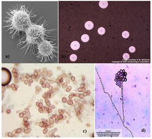
Cryptococcus neoformans: A filamentous fungus belonging to the class Tremellomycetes.
Cladosporium sphaerospermum: Member of the class Dothideomycetes, C. sphaerospermum is one of the major species observed to inhabit the destroyed reactor at Chernobyl [1].
Wangiella dermatitidis: A dematiaceous (dark-pigmented) fungus that naturally inhabits soil and plant material, this species has been occasionally associated with human disease [11].
Confirming Radiotrophic Discovery

Zhdanova et al. (1991)
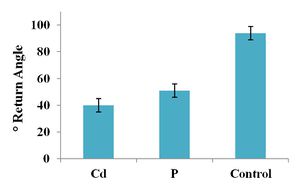
Zhdanova et al. conducted an experiment where fungal samples extracted from the remains of a Chernobyl reactor were inoculated near radioactively decaying 109Cadmium or 32Phosphorus radionuclides [1].
At the end of the growth period, 66.7% of the sample had exhibited growth in the direction towards the source of radiation independent of the direction of the food base also present in the growth vessel [1][6].
In comparison to a control group grown in a radioactivity-free environment, all the fungi growing in the presence of the decaying radionuclides exhibited greater growth, suggesting that these fungal species prefer radioactivity [1].
As well, the fungi that responded to radioactivity were commonly observed to have a high intracellular concentration of the pigment melanin [1].
Upon examining these observations, Zhdanova et al. concluded the 1991 study by suggesting that the role of melanin in fungi is analogous to that of chlorophyll in phototrophic plants with regards to the ability to obtain energy from the electromagnetic spectrum [1].
Dadachova et al. (2007)
Dadachova et al. exposed three species of fungi (C. neoformans, C. sphaerospermum, and W. dermatitidis) to heightened levels of ionizing gamma radiation from 188Rhenium and 188Tungsten [3].
Melanin-containing W. dermatitidis and C. neoformans cells exposed to ionizing gamma radiation 500 times the background levels in normal environments were found to grow at a significantly faster rate than irradiated non-melanised cells or non-irradiated melanised cells [3]. Supporting observations included higher colony-forming unit counts, more dry weight biomass, and 3-fold greater uptake of 14C-acetate by the irradiated melanised cells [3]. Similarly, heightened radiation was associated with enhanced growth in melanised C. sphaerospermum cells even under limited nutrient conditions [3].
Specifically in C. neoformans, within 20 – 40 minutes of initial exposure to increased radiation, the chemical properties of its associated melanin pigment was noted to have altered [3]. Such a change was reflected in the 3 – 4 fold increase in the rate of melanin-mediated electron transfer compared to non-irradiated cells [3]. The metabolic activity of melanin as an electron transfer agent was measured by the coupled redox reaction of NADH/ferricyanide [3].
Mechanism of Energy Harvesting
From the studies by Zhdanova et al. (1991) and Dadachova et al. (2007), the melanin found in the cell wall of radiotrophic fungi was suggested to be the prominent molecule used in harvesting usable energy from ionizing radiation.
Dadachova et al. hypothesized that ionizing radiation could change the electronic properties of melanin, such that its unpaired electrons are excited into states of higher energy [5], enabling the pigment to gain a better function in electron transfer [3]. Further experimentation by the group had yielded supportive results, where irradiated melanin was found to have manifested a 4-fold increase in its ability of reduce NADH in comparison to the non-irradiated form [3]. This would theoretically lead to more efficient energy transduction from radiation to another usable form of energy, which may enhance the growth of melanised fungi [3].
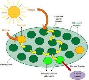
This hypothesized mechanism of energy transfer was noted to perhaps be somewhat analogous to the pigment chlorophyll in photosynthesis [1]. In phototrophs, photons from the visible range of the electromagnetic spectrum are captured by chlorophyll, whose energy is then used to generate usable chemical energy (ATP) via photophosphorylation [8]. However, whether radiotrophic fungi carry out the rest of the energy transduction process in a similar multi-step pathway as photosynthesis remains an area of active research [1].
Possible Application: Bioremediation of Radionuclides
Aside from its role as an energy transporter, melanin also appears to confer a certain degree of radioresistance in radiotrophic fungi [9]. This has led to the proposal that these fungal species may have an advanced ability, as in turn conferred by their radioresistance, to absorb and break down radioactive compounds in the environment, which may be the result of contamination by industrial effluent [9].
References
(1) Dadachova, E. and Casadevall, A. “Ionizing Radiation: how fungi cope, adapt, and exploit with the help of melanin.” Curr Opin Microbiol, 2008, DOI:10.1016/j.mib.2008.09.013
(2) Robinson, C.H. “Cold adaptation in Arctic and Antarctic fungi.” New Phytologist, 2001, DOI: 10.1046/j.1469-8137.2001.00177.x
(3) Dadachova, E., Bryan, R.A., Huang, X., Moadel, T., Schweitzer, A.D., Aisen, P., Nosanchuk, J.D., and Casadevall, A. “Ionizing Radiation Changes the Electronic Properties of Melanin and Enhances the Growth of Melanized Fungi.” PLoS ONE, 2007, DOI:10.1371/journal.pone.0000457
(4) Belozerskaya, T., Aslanidi, K., Ivanova, A., Gessler, N, Egorova, Karpenko, Yu., and Olishevskaya, S. “Characteristics of Extremophylic Fungi from Chernobyl Nuclear Power Plant.” Current Research, Technology and Education Topics in Applied Microbiology and Microbial Biotechnology. A. Mendez-Vilas (Ed.) Vol. 1, Formatex Research Centre, p88-94.
(5) Castelvecchi, D. “Dark Power: Pigment seems to put radiation to good use.” Science News, 2007, DOI:10.1002/scin.2007.5591712106
(6) Zhdanova, N., Lashkom T., Vasiliveskaya, A., Bosisyuk, L., Sinyaveskaya O., Gavrilyuk, V., and Muzale, P. “Interaction of soil micromycetes with ‘hot’ particles in a model system.” Microbiologichny Zhurnal, 1991. Abstract accessed from http://www.ncbi.nlm.nih.gov/pubmed/1753889
(7) Ionizing Radiation. Retrieved October 26, 2012, from http://www.who.int/ionizing_radiation/about/what_is_ir/en/index.html
(8) Krause, G.H., and Weis, E. “Chlorophyll Fluorescence and Photosynthesis: The Basics.” Annual Review of Plant Physiology and Plant Molecular Biology, 1991, DOI:10.1146/annurev.pp.42.060191.001525
(9) Dighton, J., Tugay, T., and Zhdanova, N. “Fungi and ionizing radiation from radionuclides.” FEMS Microbiology Letters, 2008, DOI:10.1111/j.1574-6968.2008.01076.x
(10) Dion, P., and Nautiyal, C.S., (eds). Microbiology of Extreme Soils. Heidelberg, DE: Springer-Verlag Berlin Heidelberg, 2008.
(11) Hohl, P.E., Preston-Holley Jr., H., Prevost, E., Ajello, L., and Padbye, A.A. “Infections Due to Wangiella dermatitidis in Humans: Report of the First Documented Case from the United States and a Review of the Literature.” Clin Infect Dis, 1983, DOI:10.1093/clinids/5.5854

