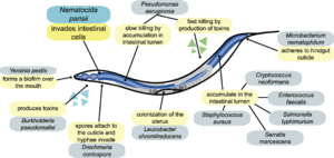
Nematocida parisii: Difference between revisions
No edit summary |
|||
| Line 40: | Line 40: | ||
==Current Research on <i>N. parisii</i>== | ==Current Research on <i>N. parisii</i>== | ||
<br>Include some current research in each topic, with at least one figure showing data.<br> | <br>Include some current research in each topic, with at least one figure showing data.<br> | ||
Revision as of 19:19, 19 April 2014
Introduction to Microsporidia

A) Shows a dormant spore containing a polar filament (black), nucleus (gray), polaroplast and posterior vacuole.
B) The posterior vacuole swells with water and ruptures the anchoring disk, allowing the polar filament to emerge through through spore cell wall.
C) Polar filament continues to to outward and evert.
D) Polar filament is fully everted and becomes a polar tube. Sporoplasm is squeezed into the polar tube.
E) Sporoplasm is moving through the polar tube.
F) Entire sporoplasm emerges from the polar tube while bound to the new membrane
Image source: http://www.annualreviews.org/doi/pdf/10.1146/annurev.micro.56.012302.160854 [1]
At right is a sample image insertion. It works for any image uploaded anywhere to MicrobeWiki. The insertion code consists of:
Double brackets: [[
Filename: PHIL_1181_lores.jpg
Thumbnail status: |thumb|
Pixel size: |300px|
Placement on page: |right|
Legend/credit: Electron micrograph of the Ebola Zaire virus. This was the first photo ever taken of the virus, on 10/13/1976. By Dr. F.A. Murphy, now at U.C. Davis, then at the CDC.
Closed double brackets: ]]
Other examples:
Bold
Italic
Subscript: H2O
Superscript: Fe3+
Background on Nematocida parisii

Image source: http://www.plosbiology.org/article/fetchObject.action?uri=info%3Adoi%2F10.1371%2Fjournal.pbio.1000005&representation=PDF [3]
Include some current research in each topic, with at least one figure showing data.
Infection and Spread of N. parisii in C. elegans
Cell shape and Metabolism
Include some current research in each topic, with at least one figure showing data.
Current Research on N. parisii
Include some current research in each topic, with at least one figure showing data.

A)
B)
C)
D)
E)
F)
Image Source: http://www.plosbiology.org/article/fetchObject.action?uri=info%3Adoi%2F10.1371%2Fjournal.pbio.0060309&representation=PDF[4]
Conclusion
Overall paper length should be 3,000 words, with at least 3 figures.
References
[8] Moretto, M.M., Khan, I.A., and Weiss, L.M. "Gastrointestinal Cell Mediated Immunity and the Microsporidia". "PLoS Pathogens". 2012. Volume 8 Issue 7. p. 1-4.
Edited by student of Joan Slonczewski for BIOL 238 Microbiology, 2014, Kenyon College.
