Nematocida parisii: Difference between revisions
| Line 32: | Line 32: | ||
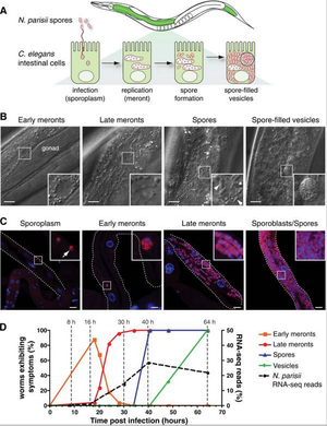
<br><br>[[Image: He3.jpg|thumb|300px|right|Comparison of infection symptoms in different. | <br><br>[[Image: He3.jpg|thumb|300px|right|Comparison of infection symptoms in different. | ||
<br>A) | |||
<br>B) | |||
<br>C) | |||
<br>D) | |||
<br>E) | |||
<br>F) <br> <br>Image Source: http://www.plosbiology.org/article/fetchObject.action?uri=info%3Adoi%2F10.1371%2Fjournal.pbio.0060309&representation=PDF[3]]] | |||
==Conclusion== | ==Conclusion== | ||
Revision as of 02:06, 17 April 2014
Introduction to Microsporidia
At right is a sample image insertion. It works for any image uploaded anywhere to MicrobeWiki. The insertion code consists of:
Double brackets: [[
Filename: PHIL_1181_lores.jpg
Thumbnail status: |thumb|
Pixel size: |300px|
Placement on page: |right|
Legend/credit: Electron micrograph of the Ebola Zaire virus. This was the first photo ever taken of the virus, on 10/13/1976. By Dr. F.A. Murphy, now at U.C. Davis, then at the CDC.
Closed double brackets: ]]
Other examples:
Bold
Italic
Subscript: H2O
Superscript: Fe3+
Background on Nematocida parisii
Include some current research in each topic, with at least one figure showing data.
Cell shape and Metabolism
Include some current research in each topic, with at least one figure showing data.
Pathogenesis in Caenorhabditis elegans
Include some current research in each topic, with at least one figure showing data.

A)
B)
C)
D)
E)
F)
Image Source: http://www.plosbiology.org/article/fetchObject.action?uri=info%3Adoi%2F10.1371%2Fjournal.pbio.0060309&representation=PDF[3]
Conclusion
Overall paper length should be 3,000 words, with at least 3 figures.
References
[4]
[5]
[6]
Edited by student of Joan Slonczewski for BIOL 238 Microbiology, 2014, Kenyon College.


