Nematocida parisii: Difference between revisions
| Line 40: | Line 40: | ||
<br><br>Image source: http://genome.cshlp.org/content/22/12/2478.full.pdf+html [3]]] | <br><br>Image source: http://genome.cshlp.org/content/22/12/2478.full.pdf+html [3]]] | ||
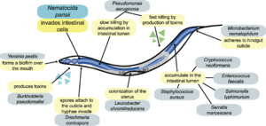
N. parisii is transmitted horizontally from animal to animal [6]. N. parisii enters the intestinal region of C. elegans through either oral consumption or rectal entrance. It infects C. elegans intestinal cells during its transmissible spore form, outlined in Figure 3A [7]. Following the identical spore germination outlined in microsporidia, N. parisii spores invert their polar filament to form a polar tube. The polar tube breaks through the cell wall and cell membrane of the spore and launches itself through the cell membrane of a nearby host cell, in this case the C. elegans intestinal cell. The polar tube serves as a bridge between the spore and the intestinal cell and injects the N. parisii sporoplasm into the C. elegans intestinal cell. Sporoplasm becomes a meront and develops to generate mature spores. Figure 3B gives phase-by-phase visual of post-sporoplasm injection in C. elegans and the development of meront to how the host cell becomes filled with Ni parisii spores. Figure 3C gives a visual of the rapid spore replication during the time of replication. Notice as the N. parisii (red) take over the C. elegans intestinal cells (blue), but they do not disappear. Usually when pathogens such as viruses produce progeny in a host cell, the cell lyses and release all the progeny viruses out. However, from the stain, there is a reasonable concentration of purple in the area where intestinal cells were located, indicating the cells are still alive [5]. As it turns out, mature N. parisii spores are able to exit the host cell without causing severe damage to the cell. N. parisii is able to manipulate the host cell’s cytoskeleton to commit non-lytic escape [7]. | |||
<br>Electron microscope analysis of N. parisii infection in intestinal cells show that the presence of N. parisii in the intestinal cell results in alteration of the terminal web [6,7]. The terminal web is a cytoskeletal structure found in many polarized epithelial cells [6]. N. parisii is able to rearrange ACT-5, a specialized actin isoform that is associated with both the terminal web and microvilli of C. elegans. ACT-5 is relocalized from the apical side to the ecotopical basolacteral side of the cell [7]. The actin relocalization that occurred leave gaps in the terminal web, approximately 1 m-wide [7]. These gaps allow the N. parisii spores formed from the meront to escape the host cell and into the C. elegans lumen without causing cell lysation. Leaving the host cell alive is beneficial to N. parisii because it keeps the host alive to pass off the pathogen to another healthy host [7]. | <br>Electron microscope analysis of N. parisii infection in intestinal cells show that the presence of N. parisii in the intestinal cell results in alteration of the terminal web [6,7]. The terminal web is a cytoskeletal structure found in many polarized epithelial cells [6]. N. parisii is able to rearrange ACT-5, a specialized actin isoform that is associated with both the terminal web and microvilli of C. elegans. ACT-5 is relocalized from the apical side to the ecotopical basolacteral side of the cell [7]. The actin relocalization that occurred leave gaps in the terminal web, approximately 1 m-wide [7]. These gaps allow the N. parisii spores formed from the meront to escape the host cell and into the C. elegans lumen without causing cell lysation. Leaving the host cell alive is beneficial to N. parisii because it keeps the host alive to pass off the pathogen to another healthy host [7]. | ||
Revision as of 20:04, 19 April 2014
Introduction to Microsporidia

A) Shows a dormant spore containing a polar filament (black), nucleus (gray), polaroplast and posterior vacuole.
B) The posterior vacuole swells with water and ruptures the anchoring disk, allowing the polar filament to emerge through through spore cell wall.
C) Polar filament continues to to outward and evert.
D) Polar filament is fully everted and becomes a polar tube. Sporoplasm is squeezed into the polar tube.
E) Sporoplasm is moving through the polar tube.
F) Entire sporoplasm emerges from the polar tube while bound to the new membrane
Image source: http://www.annualreviews.org/doi/pdf/10.1146/annurev.micro.56.012302.160854 [1]
At right is a sample image insertion. It works for any image uploaded anywhere to MicrobeWiki. The insertion code consists of:
Double brackets: [[
Filename: PHIL_1181_lores.jpg
Thumbnail status: |thumb|
Pixel size: |300px|
Placement on page: |right|
Legend/credit: Electron micrograph of the Ebola Zaire virus. This was the first photo ever taken of the virus, on 10/13/1976. By Dr. F.A. Murphy, now at U.C. Davis, then at the CDC.
Closed double brackets: ]]
Other examples:
Bold
Italic
Subscript: H2O
Superscript: Fe3+
Background on Nematocida parisii

Image source: http://www.plosbiology.org/article/fetchObject.action?uri=info%3Adoi%2F10.1371%2Fjournal.pbio.1000005&representation=PDF [3]
Include some current research in each topic, with at least one figure showing data.
Infection and Spread of N. parisii in C. elegans

A) Visual of N. parisii infection of C. elegans intestinal cells (green) from sporoplasm invasion to meront formation to replication and formation of spores.
B) DIC images of dissected animals showed various stages of infection. Arrows point out large spores.
C) FISH (red) and DAPI (blue) staining of N. parisii at various stages of infection.
Image source: http://genome.cshlp.org/content/22/12/2478.full.pdf+html [3]
N. parisii is transmitted horizontally from animal to animal [6]. N. parisii enters the intestinal region of C. elegans through either oral consumption or rectal entrance. It infects C. elegans intestinal cells during its transmissible spore form, outlined in Figure 3A [7]. Following the identical spore germination outlined in microsporidia, N. parisii spores invert their polar filament to form a polar tube. The polar tube breaks through the cell wall and cell membrane of the spore and launches itself through the cell membrane of a nearby host cell, in this case the C. elegans intestinal cell. The polar tube serves as a bridge between the spore and the intestinal cell and injects the N. parisii sporoplasm into the C. elegans intestinal cell. Sporoplasm becomes a meront and develops to generate mature spores. Figure 3B gives phase-by-phase visual of post-sporoplasm injection in C. elegans and the development of meront to how the host cell becomes filled with Ni parisii spores. Figure 3C gives a visual of the rapid spore replication during the time of replication. Notice as the N. parisii (red) take over the C. elegans intestinal cells (blue), but they do not disappear. Usually when pathogens such as viruses produce progeny in a host cell, the cell lyses and release all the progeny viruses out. However, from the stain, there is a reasonable concentration of purple in the area where intestinal cells were located, indicating the cells are still alive [5]. As it turns out, mature N. parisii spores are able to exit the host cell without causing severe damage to the cell. N. parisii is able to manipulate the host cell’s cytoskeleton to commit non-lytic escape [7].
Electron microscope analysis of N. parisii infection in intestinal cells show that the presence of N. parisii in the intestinal cell results in alteration of the terminal web [6,7]. The terminal web is a cytoskeletal structure found in many polarized epithelial cells [6]. N. parisii is able to rearrange ACT-5, a specialized actin isoform that is associated with both the terminal web and microvilli of C. elegans. ACT-5 is relocalized from the apical side to the ecotopical basolacteral side of the cell [7]. The actin relocalization that occurred leave gaps in the terminal web, approximately 1 m-wide [7]. These gaps allow the N. parisii spores formed from the meront to escape the host cell and into the C. elegans lumen without causing cell lysation. Leaving the host cell alive is beneficial to N. parisii because it keeps the host alive to pass off the pathogen to another healthy host [7].
Cell shape and Metabolism
Include some current research in each topic, with at least one figure showing data.
Current Research on N. parisii
Include some current research in each topic, with at least one figure showing data.

A)
B)
C)
D)
E)
F)
Image Source: http://www.plosbiology.org/article/fetchObject.action?uri=info%3Adoi%2F10.1371%2Fjournal.pbio.0060309&representation=PDF[4]
Conclusion
Overall paper length should be 3,000 words, with at least 3 figures.
References
[8] Moretto, M.M., Khan, I.A., and Weiss, L.M. "Gastrointestinal Cell Mediated Immunity and the Microsporidia". "PLoS Pathogens". 2012. Volume 8 Issue 7. p. 1-4.
Edited by student of Joan Slonczewski for BIOL 238 Microbiology, 2014, Kenyon College.
