File:3AD2E51A-D488-41AD-AC2A-92ACC6052754 4 5005 c.jpeg
3AD2E51A-D488-41AD-AC2A-92ACC6052754_4_5005_c.jpeg (544 × 210 pixels, file size: 63 KB, MIME type: image/jpeg)
Summary
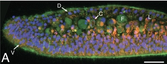
This photo shows Trichoplax adhaerens as prepared by a microscope in a cross-section. The different stains allow different parts of the cell to be seen. In light green you see the autofluorescence. The bright green at the bottom shows filamentous actin that are present within epithelial cells. The blue-violet color depicts nuclei in epithelial cells. The reddish orange shows lipophil cells. Lastly, the pale green indicates the fiber cell bodies, more specifically their auto-fluorescent granules. Credit to Carolyn Smith and Thomas Reese.
File history
Click on a date/time to view the file as it appeared at that time.
| Date/Time | Thumbnail | Dimensions | User | Comment | |
|---|---|---|---|---|---|
| current | 00:33, 13 December 2022 | 544 × 210 (63 KB) | Map1934 (talk | contribs) | This photo shows ''Trichoplax adhaerens'' as prepared by a microscope in a cross-section. The different stains allow different parts of the cell to be seen. In light green you see the autofluorescence. The bright green at the bottom shows filamentous actin that are present within epithelial cells. The blue-violet color depicts nuclei in epithelial cells. The reddish orange shows lipophil cells. Lastly, the pale green indicates the fiber cell bodies, more specifically their auto-fluorescent gr... |
You cannot overwrite this file.
File usage
The following page uses this file:
