File:Cell envelope.jpg
From MicrobeWiki, the student-edited microbiology resource
No higher resolution available.
Cell_envelope.jpg (600 × 470 pixels, file size: 103 KB, MIME type: image/jpeg)
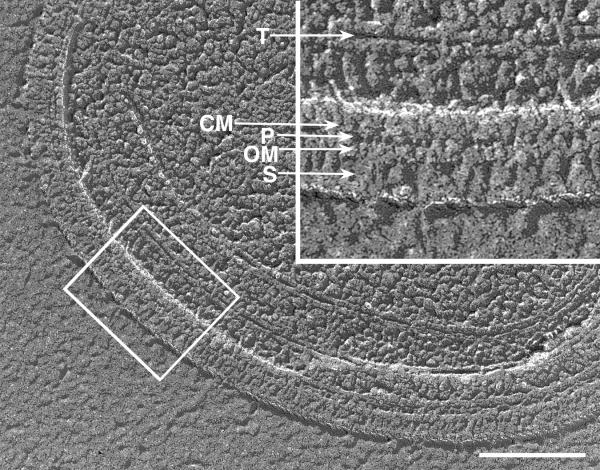
Membrane structure of Synechococcus (strain WH8113) The inset corresponds to the outlined section of cell envelope comprising cell membrane (CM), peptidoglycan layer (P), outer membrane (OM), and surface layer (S). A thylakoid layer (T) is also indicated. This picture was published in 2001 by Aravinthan DT Samuel, Jennifer D. Petersen and Thomas S. Reese.
File history
Click on a date/time to view the file as it appeared at that time.
| Date/Time | Thumbnail | Dimensions | User | Comment | |
|---|---|---|---|---|---|
| current | 09:13, 12 March 2014 | | 600 × 470 (103 KB) | Cyoung9350 (talk | contribs) | Membrane structure of Synechococcus (strain WH8113) The inset corresponds to the outlined section of cell envelope comprising cell membrane (CM), peptidoglycan layer (P), outer membrane (OM), and surface layer (S). A thylakoid layer (T) is also indicat... |
You cannot overwrite this file.
File usage
The following page uses this file:
