File:Alcanivorax bork.jpg: Difference between revisions
From MicrobeWiki, the student-edited microbiology resource
(Mhsu23 uploaded a new version of "File:Alcanivorax bork.jpg": Reverted to version as of 16:12, 13 May 2015) |
No edit summary |
||
| Line 1: | Line 1: | ||
Transmission electron micrographs of “Candidatus Scalindua sp.” enriched in the MBR, showing the cells which were about to be divided. All cells in | |||
panels A and B were divided into three separate compartments by individual membranes: the paryphoplasm, the riboplasm, and the anammoxosome compartments. | |||
In panel A, all cells were occupied by the voluminous anammoxosome. In panel B, the white arrows indicate condensed and electron-dense particles. Scale | |||
bars, 500 nm. | |||
Latest revision as of 16:23, 13 May 2015
Transmission electron micrographs of “Candidatus Scalindua sp.” enriched in the MBR, showing the cells which were about to be divided. All cells in panels A and B were divided into three separate compartments by individual membranes: the paryphoplasm, the riboplasm, and the anammoxosome compartments. In panel A, all cells were occupied by the voluminous anammoxosome. In panel B, the white arrows indicate condensed and electron-dense particles. Scale bars, 500 nm.
File history
Click on a date/time to view the file as it appeared at that time.
| Date/Time | Thumbnail | Dimensions | User | Comment | |
|---|---|---|---|---|---|
| current | 16:22, 13 May 2015 |  | 754 × 507 (227 KB) | Mhsu23 (talk | contribs) | Reverted to version as of 16:12, 13 May 2015 |
| 16:13, 13 May 2015 | 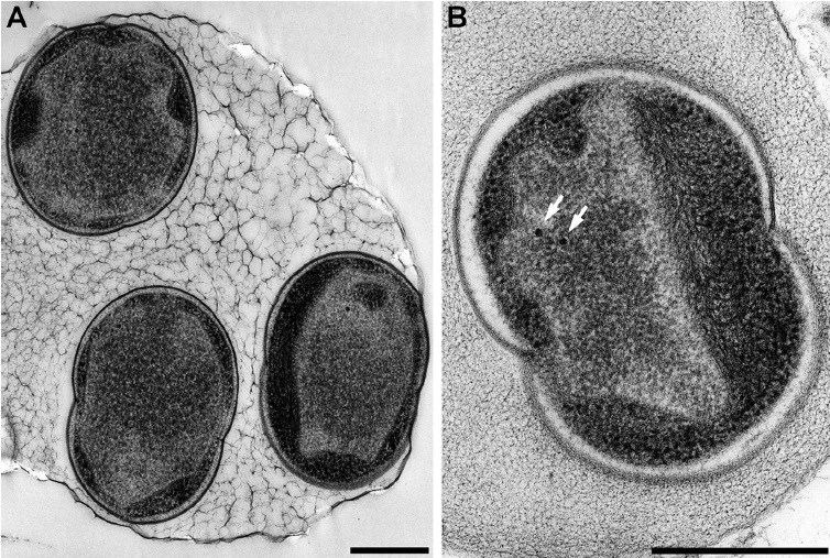 | 754 × 507 (227 KB) | Mhsu23 (talk | contribs) | ||
| 16:12, 13 May 2015 |  | 754 × 507 (227 KB) | Mhsu23 (talk | contribs) | ||
| 01:32, 14 December 2012 | 800 × 319 (45 KB) | Pawan dhaliwal89 (talk | contribs) |
You cannot overwrite this file.
File usage
The following 3 files are duplicates of this file (more details):
The following 5 pages use this file:
