Brachyspira hyodysenteriae: Difference between revisions
No edit summary |
No edit summary |
||
| Line 1: | Line 1: | ||
{{MooreL}} | |||
{{Uncurated}} | {{Uncurated}} | ||
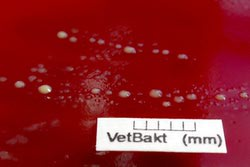
[[File:bh-1.jpeg|250px|thumb|right|''Brachyspira hyodysenteriae'' [http://www.brachyspira.se/brachyspira/popups/image.php?imgtable=brachy_images1&imgid=1&image=0 Image from Karl-Erik Johansson and Therese Råsbäck]]] | [[File:bh-1.jpeg|250px|thumb|right|''Brachyspira hyodysenteriae'' [http://www.brachyspira.se/brachyspira/popups/image.php?imgtable=brachy_images1&imgid=1&image=0 Image from Karl-Erik Johansson and Therese Råsbäck]]] | ||
''Brachyspira hyodysenteriae'' | ''Brachyspira hyodysenteriae'' | ||
Revision as of 19:19, 5 February 2016

Brachyspira hyodysenteriae
Classification
Higher order taxa
Domain:Bacteria Phylum: Spirochaetes Class: Spirochaetia Order: Spirochaetales Family: Brachyspiraceae Genus:Brachyspira[6]
Species
hyodysenteriae
NCBI: Taxonomy Brachyspira hyodysenteriae
Description and significance
B. hyodysenteriae is able to survive transiently primarily in moist feces from infected pigs which allows for an increase in the spread of the bacteria among pigs via digestion of infected feces.[2] Once a pig is infected with B. hyodysenteriae it will most likely develop swine dysentery (SD). Swine dysentery can lead to stunted growth rates and death which can be extremely costly when the swine are raised for food purposes.[1] There are very few drugs commercially available to combat the costly side effects of SD due to its insensitivity and resistance to macrolide, lincosamide, and pleuromutilin classes of antibiotics typically used to treat SD.[5]
Genome structure
B. hyodysenteriae has one circular chromosome consisting of 3,000,694 base pairs. There is also a circular plasmid that consists of 35,940 base pairs. The genome contains 2,669 open reading frames with 2,122 being putative protein-coding sequences.[2]
Cell and colony structure

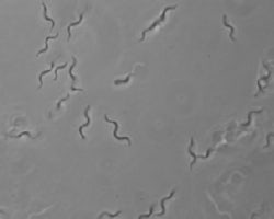
"B. hyodysenteriae" is a gram negative bacterium with a loosely coiled, helical shape. These microbes have flagella in the periplasmic space, which is typical of spirochaetes, to assist in motility which is necessary for colony formation in the large intestine.[3]
Metabolism
B. hyodysenteriae is a facultative anaerobe which is oxygen tolerant due to its NADH oxidase activity.[2] It is a chemoorganotroph with acetate, butyrate, H2, and CO2 being major end products resulting from glucose metabolism. B. hyodysenteriae is readily grown on tryptic soy agar plates or in brain heart infusion salt or Kunkle’s broth at 38-40°C with a growth yield of ~109 cells/ml and a doubling time of 1-5 hours. [5]
Ecology
Usually found in the large intestine of a pig or living transiently in moist feces excreted from an infected animal.[2] B. hyodysenteriae is known to mostly affect piglets but has also been found to cause disease in chickens and birds as well.[7]
Pathology
This pathogenic bacteria colonizes in the large intestine of pigs causing dysentery. [2] The complete haemolysis of the red blood cells contributes is the reason B. hyodysenteriae is so highly pathogenic.[1]
References
1. Novotna M &Skardova O. Brachyspira hyodysenteriae:detection, identification and antibiotic susceptibility. Vet. Med.-Czech, 47,2002(4):104-109.
2. Bellgard MI, Wanchanthuek P, La T, Ryan K, Moolhuijzen P, et al. (2009) Genome Sequence of the Pathogenic Intestinal Spirochete Brachyspira hyodysenteriae Reveals Adaptations to Its Lifestyle in the Porcine Large Intestine. PLoS ONE 4(3): e4641. doi:10.1371/journal.pone.0004641
3. Chunhao Li, Charles W. Wolgemuth, Michael Marko, David G. Morgan, Nyles W. Charon. J Bacteriol. 2008 August; Genetic Analysis of Spirochete Flagellin Proteins and Their Inolvement in Motility, Filament Assembly Morphology. 190(16): 5607–5615. Published online 2008 June 13. doi: 10.1128/JB.00319-08
4. M. Karlsson, A. Aspán, A. Landén, and A. Franklin Further characterization of porcine Brachyspira hyodysenteriae isolates with decreased susceptibility to tiamulin J Med Microbiol April 2004 53:281-285; doi:10.1099/jmm.0.05395-0
5. Paster B.J. 2010. Phylum XV Spirochaetes, Garrity and Holt 2001. Bergey’s Manual of Systematic Bacteriology 533-537.
6. http://www.ncbi.nlm.nih.gov/Taxonomy/Browser/wwwtax.cgi
7. Anneke Feberwee, David J. Hampson, Nyree D. Phillips, Tom La, Harold M. J. F. van der Heijden, Gerard J. Wellenberg, R. Marius Dwars, Wil J. M. Landman. Identification of Brachyspira hyodysenteriae and Other Pathogenic Brachyspira Species in Chickens from Laying Flocks with Diarrhea or Reduced Production or Both. J Clin Microbiol. 2008 February; 46(2): 593–600. doi: 10.1128/JCM.01829-07
Edited by Chelsea Douglass student of Dr. Lisa Moore of the University of Southern Maine
