Genus S and J: Difference between revisions
No edit summary |
|||
| (28 intermediate revisions by the same user not shown) | |||
| Line 1: | Line 1: | ||
{{Uncurated}} | {{Uncurated}} | ||
[[File:Pseudomonasaeruginosa.jpg|frame|right|<i>Pseudomonas aeruginosa<i> http://pin.it/eKZkdDj ]] | |||
==Classification== | ==Classification== | ||
<p>Kingdom: Bacteria | |||
< | <p>Phylum: Proteobacteria | ||
<p>Class: Gammaproteobacteria | |||
<p>Order: Pseudomonadales | |||
Phylum: Proteobacteria | <p>Family: Pseudomonadaceae | ||
Class: Gammaproteobacteria | <p>Genus: Pseudomonas | ||
Order: Pseudomonadales | <p>Species: <b><i> P. aeruginosa</i></b></p> | ||
Family: Pseudomonadaceae | |||
Genus: Pseudomonas | |||
Species: <i> P. aeruginosa<i> <b> | |||
===Species=== | ===Species=== | ||
{| | {| | ||
| height="10" bgcolor="#FFDF95" | | | height="10" bgcolor="#FFDF95" | | ||
| Line 21: | Line 17: | ||
|} | |} | ||
''Pseudomonas aeruginosa'' | ''<i>Pseudomonas aeruginosa<i>'' | ||
Taxonomy ID: 1454219 | <p>Taxonomy ID: 1454219</p> | ||
Inherited blast name: g-proteobacteria | <p>Inherited blast name: g-proteobacteria</p> | ||
==Habitat Information == | ==Habitat Information == | ||
The soil was obtained on September 8, 2016 during a clear night around 9:30 PM. There was an estimate of 85% humidity and the temperature was around 85 degrees Fahrenheit with 0 mph wind. I browsed around my apartment complex came across a grassy area near one of the pools right behind my apartment unit. The grass was not recently watered and there had been no recent rainfall prior to collecting the soil sample. | |||
<p>Map Unit Name: Tarrant and Speck soils, 0 to 2 percent slopes</p> | |||
== | ==Description and Significance== | ||
*Interesting features of cell structure; how it gains energy; what important molecules it produces. | |||
===Cell Structure, Metabolism, and Life Cycle=== | |||
<i>Pseudomonas aeruginosa<i> is a Gram-negative and bacillus (rod-shaped) bacterium. Gram-negative indicates that it has an inner layer of peptidoglycan in its cell wall, as well as an outer layer of lipoprotein and lipopolysaccharides. Its colonial color is brown or metallic sheen, with a blue-green extracellular pigment. [[File:MasterPlatewithDisk.jpg|thumb|left|<i>P. Aeruginosa<i> Master Plate]] Its colonial morphology is small, entire, convex, and mucoid. <i>P. aeruginosa<i> has a blue-green appearance when cultured, which stems from the latin word <i>"aeruginosa"<i> meaning "copper rust". However, from our culture of patch number 5, this microorganism appeared cloudy white. | |||
< | <i>P. aeruginosa<i> is a facultative anaerobe, which indicates that it can produce ATP by aerobic respiration in the presence of oxygen or by anaerobic respiration in absence of oxygen. | ||
This microorganism has a tendency to form biofilms, which are substances that help organisms adhere to various surfaces. Once biofilms are formed, they are pretty difficult to destroy, which improves the efficiency of <i>Pseudomonas Aeruginosa<i> in pathogenic activity. | |||
===Significance=== | |||
<p><i>P. aeruginosa<i> is considered one of the most common gram-negative infections in hospital settings, especially in settings consisting of weaker immune systems. [3] Exposure to this microorganism can be contracted all over the body, and result in multiple bodily effects, such as pneumonia in the respiratory tract and osteomyelitis in the bones and joints. This bacteria can be found in water, which can lead to eye and ear infections by direct contact with this microorganism. It can also cause mild illnesses and rashes in both children and adults. Therefore, it is essential to maintain clean environments for patients and healthcare providers because <i>P. aeruginosa<i> can easily spread from patient to patient [3]. These infections are generally treatable with antibiotics, but are becoming more difficult to treat overtime due to antibiotic resistance.</p> | |||
==Genome Structure== | |||
We identified the organism using PCR and gene sequencing of its rRNA genes below.<br> | |||
<p>[[File:PCR Sequencing.png|frame|center|float|360x320px|Forward PCR Sequence (left) and Reverse PCR Sequence (right)]]</p> | |||
This organism has one circular chromosome and divides by binary fission. | |||
==Physiology and Pathogenesis== | |||
<p><i>P. aeruginosa<i> is an opportunistic pathogenic microorganism. It is the most common cause of burn injuries and can spread through contaminants, especially in health care settings [2]. Within low phosphate levels, it can induce release of lethal toxins to the intestinal tract and cause severe damage to the host cells. This occurrence can be counteracted by exposing patients to an excessive level of phosphate.</p> | |||
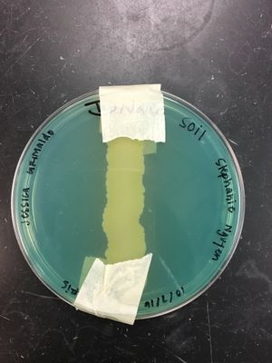
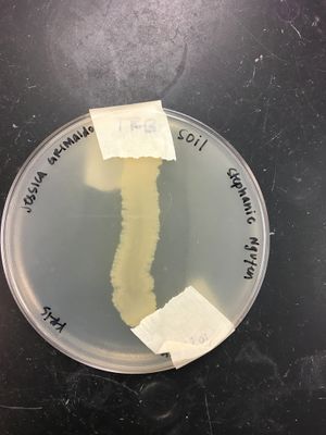
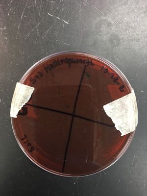
Some laboratory tests were performed in order to lead to the correctly identified microorganism within the tested soil. | |||
A chart and pictures of performed stains and experiments are provided below to summarize the results discovered in lab. | |||
<p>[[File:Laboratory Test Results.png|frame|left|600x520px|Laboratory Test Results]]</p> | |||
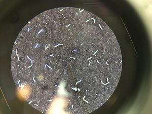
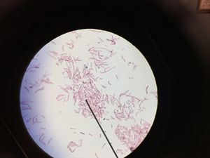
<p>[[File:Gram stain negative.jpg|thumb|right|Gram Stain - Negative]]</p> | |||
<p>[[File:Capsule stain.jpg|thumb|right|Capsule Stain]]</p> | |||
<p>[[File:Endospore stain.jpg|thumb|right|Endospore Stain]]</p> | |||
<p>[[File:Starch Hydrolysis.jpg|thumb|right|Starch Hydrolysis]]</p> | |||
<p>[[File:Casein Hydrolysis Test.jpg|thumb|right|Casein Hydrolysis]]</p> | |||
<p>[[File:DNA Hydrolysis.jpg|thumb|left|DNA Hydrolysis]]</p> | |||
<p>[[File:Lipid Hydrolysis.jpg|thumb|left|Lipid Hydrolysis]]</p> | |||
<p>[[File:Eosin Methylene Blue.jpg|thumb|left|Eosin Methylene Blue Agar]]</p> | |||
<p>[[File:Hektoin Enteric Agar.jpg|thumb|left|Hektoen Enteric Agar]]</p> | |||
<p>[[File:MacConkey Agar.jpg|thumb|left|MacConkey Agar]]</p> | |||
<p>[[File:Decarboxylation.jpg|thumb|left|Decarboxylation]]</p> | |||
<p>[[File:Phenylalanine Deaminase Test.jpg|thumb|left|Phenylalanine Deaminase]]</p> | |||
<p>[[File:Blood Agar.jpg|thumb|left|Blood Agar]]</p> | |||
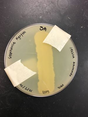
<p>[[File:Mannitol Salt Agar.jpg|thumb|left|Mannitol Salt Agar]]</p> | |||
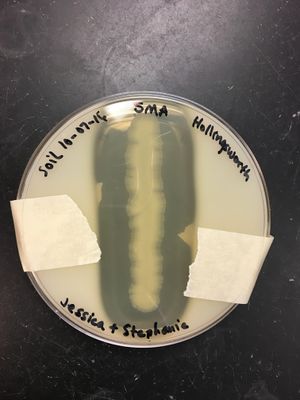
<p>[[File:Phenylethyl Alcohol Agar.jpg|thumb|left|Phenylethyl Alcohol Agar]]</p> | |||
<p>[[File:Antimicrobial Agents.jpg|thumb|left|Antimicrobial Sensitivity]]</p> | |||
<p>[[File:Disinfectants.jpg|thumb|left|Disinfectant Sensitivity]]</p> | |||
P. aeruginosa causes 10-20% of nosocomial infections and is especially pathogenic to | <br><b><i>P. aeruginosa:<i></b></br> | ||
<br> - <b>Gram-negative:</b> This means it has an inner layer of peptidoglycan in its cell wall as well as an outer layer of lipoprotein and lipopolysaccharides. </br> | |||
<br> - <b>Endospore negative:</b> It does not form spores when the environment is unfavorable. Therefore, it does not produce spores that increase pathogenicity that make it harder to destroy in the body without also harming good cells. </br> | |||
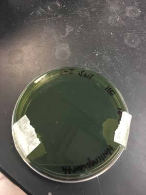
<br> - Produces the enzyme <b>deaminase</b>, which removes the amine group from the amino acid phenylalanine and releases it as ammonia, producing phenylpyruvic acid as a result. When 10% ferric chloride is added to this medium, the presence of phenylpyruvic acid causes the media to turn dark green, a positive test result for deaminase <i>(see picture below)<i></br> | |||
<br> - Causes 10-20% of <b>nosocomial infections</b> (e.g., urinary tract infections and respiratory pneumonia) and is especially pathogenic to immune-suppressed patients such as burn victims, organ transplants, cystic fibrosis patients, and those who have long stays in hospital settings. An infection can cause meningitis, endocarditis, septicemia and more. [1]</br> | |||
<br> - Can be treated with <b>aminoglycosides and penicillins</b>. However, prompt attention is <u>vital<u>.</br> | |||
==References== | ==References== | ||
[ | [1] [https://www.ncbi.nlm.nih.gov/pubmed/6405475 Bodey GP, Bolivar R, Fainstein V, Jadeja L. "'Infections caused by Pseudomonas aeruginosa'". ''Reviews of Infectious Diseases''. 1983. Volume 5. p. 279-313.]<br> | ||
[2] [http://emedicine.medscape.com/article/226748-overview Friedrich, Marcus. "Pseudomonas Aeruginosa Infections." Pseudomonas Aeruginosa Infections: Practice Essentials, Background, Pathophysiology. Medscape, 5 Dec. 2016. Web. 06 Dec. 2016.]<br> | |||
[3] [http://www.cdc.gov/hai/organisms/pseudomonas.html "Pseudomonas Aeruginosa in Healthcare Settings." Centers for Disease Control and Prevention. Centers for Disease Control and Prevention, 07 May 2014. Web. 06 Dec. 2016.]<br> | |||
==Author== | ==Author== | ||
Latest revision as of 20:26, 9 December 2016
Classification
Kingdom: Bacteria
Phylum: Proteobacteria
Class: Gammaproteobacteria
Order: Pseudomonadales
Family: Pseudomonadaceae
Genus: Pseudomonas
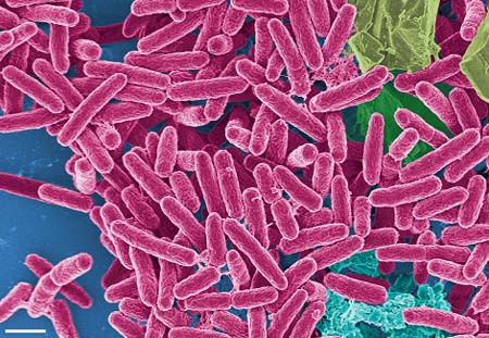
Species: P. aeruginosa
Species
|
NCBI: Taxonomy |
Pseudomonas aeruginosa
Taxonomy ID: 1454219
Inherited blast name: g-proteobacteria
Habitat Information
The soil was obtained on September 8, 2016 during a clear night around 9:30 PM. There was an estimate of 85% humidity and the temperature was around 85 degrees Fahrenheit with 0 mph wind. I browsed around my apartment complex came across a grassy area near one of the pools right behind my apartment unit. The grass was not recently watered and there had been no recent rainfall prior to collecting the soil sample.
Map Unit Name: Tarrant and Speck soils, 0 to 2 percent slopes
Description and Significance
- Interesting features of cell structure; how it gains energy; what important molecules it produces.
Cell Structure, Metabolism, and Life Cycle
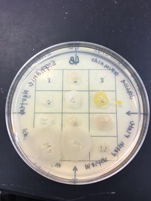
Pseudomonas aeruginosa is a Gram-negative and bacillus (rod-shaped) bacterium. Gram-negative indicates that it has an inner layer of peptidoglycan in its cell wall, as well as an outer layer of lipoprotein and lipopolysaccharides. Its colonial color is brown or metallic sheen, with a blue-green extracellular pigment.
Its colonial morphology is small, entire, convex, and mucoid. P. aeruginosa has a blue-green appearance when cultured, which stems from the latin word "aeruginosa" meaning "copper rust". However, from our culture of patch number 5, this microorganism appeared cloudy white.
P. aeruginosa is a facultative anaerobe, which indicates that it can produce ATP by aerobic respiration in the presence of oxygen or by anaerobic respiration in absence of oxygen.
This microorganism has a tendency to form biofilms, which are substances that help organisms adhere to various surfaces. Once biofilms are formed, they are pretty difficult to destroy, which improves the efficiency of Pseudomonas Aeruginosa in pathogenic activity.
Significance
P. aeruginosa is considered one of the most common gram-negative infections in hospital settings, especially in settings consisting of weaker immune systems. [3] Exposure to this microorganism can be contracted all over the body, and result in multiple bodily effects, such as pneumonia in the respiratory tract and osteomyelitis in the bones and joints. This bacteria can be found in water, which can lead to eye and ear infections by direct contact with this microorganism. It can also cause mild illnesses and rashes in both children and adults. Therefore, it is essential to maintain clean environments for patients and healthcare providers because P. aeruginosa can easily spread from patient to patient [3]. These infections are generally treatable with antibiotics, but are becoming more difficult to treat overtime due to antibiotic resistance.
Genome Structure
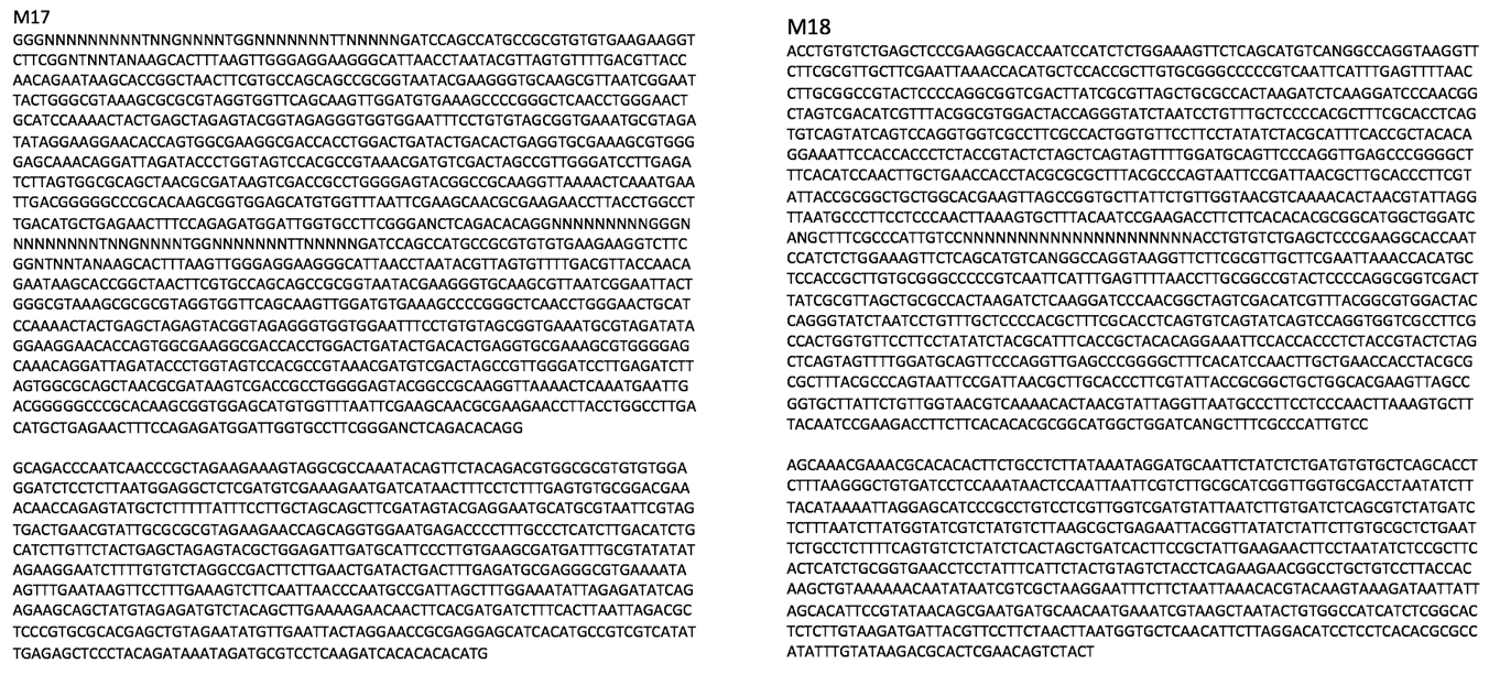
We identified the organism using PCR and gene sequencing of its rRNA genes below.
This organism has one circular chromosome and divides by binary fission.
Physiology and Pathogenesis
P. aeruginosa is an opportunistic pathogenic microorganism. It is the most common cause of burn injuries and can spread through contaminants, especially in health care settings [2]. Within low phosphate levels, it can induce release of lethal toxins to the intestinal tract and cause severe damage to the host cells. This occurrence can be counteracted by exposing patients to an excessive level of phosphate.
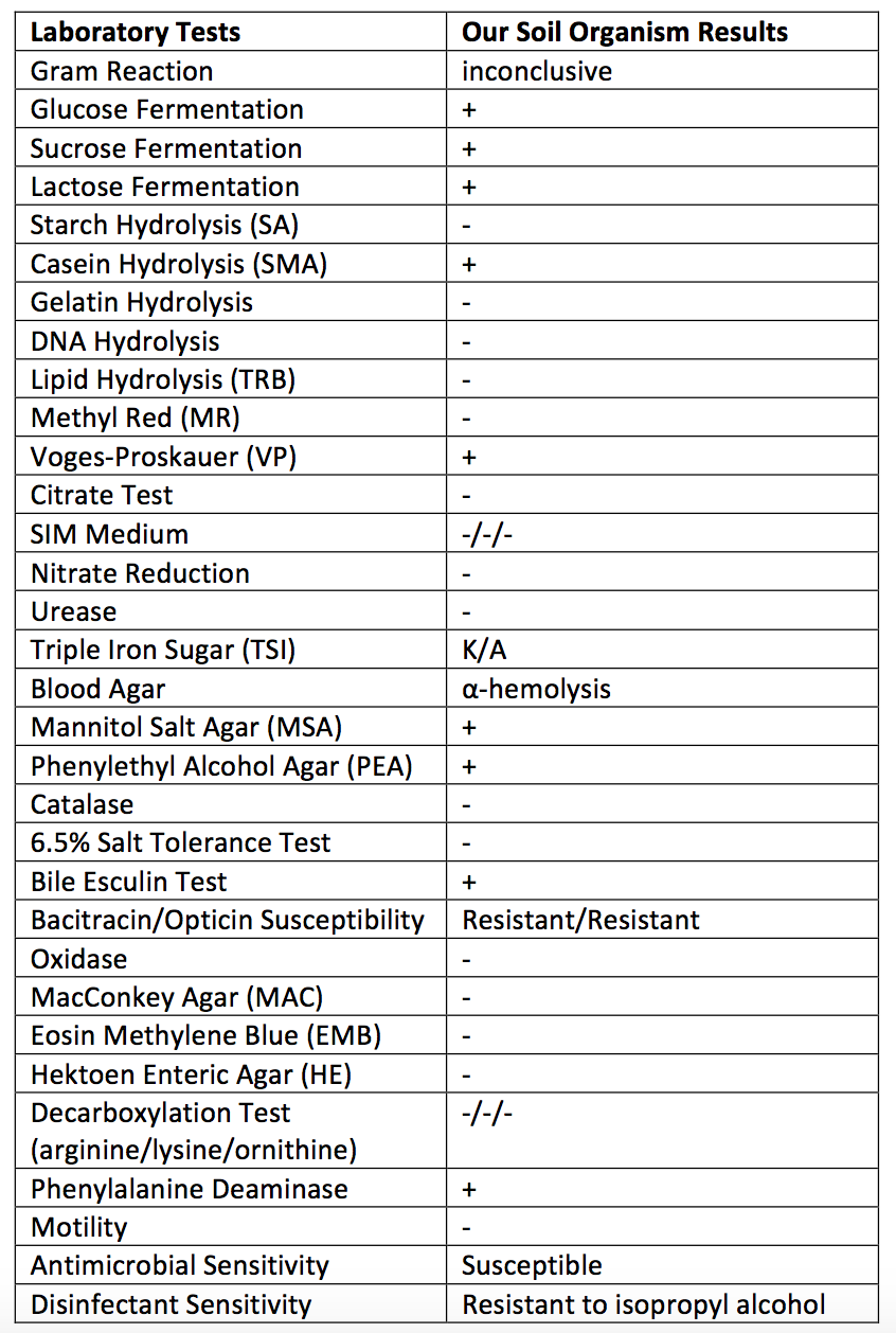
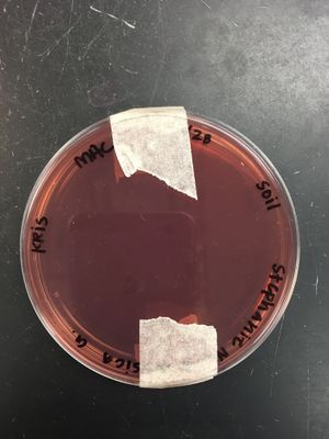
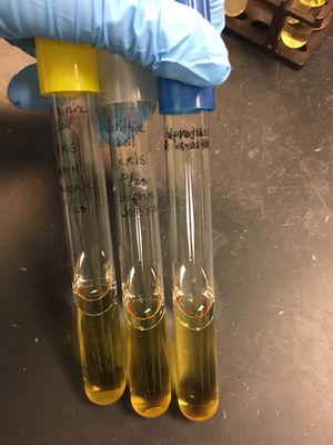
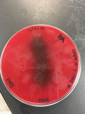
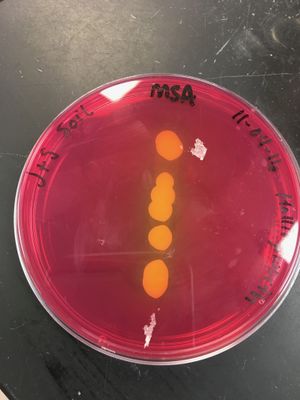
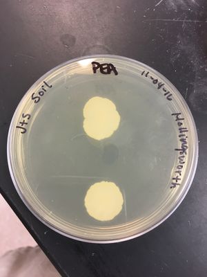
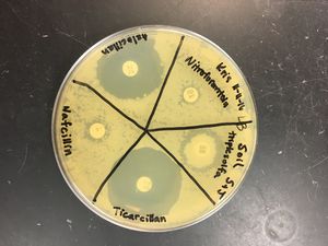
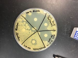
Some laboratory tests were performed in order to lead to the correctly identified microorganism within the tested soil.
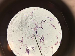
A chart and pictures of performed stains and experiments are provided below to summarize the results discovered in lab.
P. aeruginosa:
- Gram-negative: This means it has an inner layer of peptidoglycan in its cell wall as well as an outer layer of lipoprotein and lipopolysaccharides.
- Endospore negative: It does not form spores when the environment is unfavorable. Therefore, it does not produce spores that increase pathogenicity that make it harder to destroy in the body without also harming good cells.
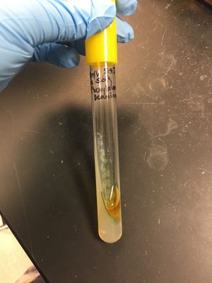
- Produces the enzyme deaminase, which removes the amine group from the amino acid phenylalanine and releases it as ammonia, producing phenylpyruvic acid as a result. When 10% ferric chloride is added to this medium, the presence of phenylpyruvic acid causes the media to turn dark green, a positive test result for deaminase (see picture below)
- Causes 10-20% of nosocomial infections (e.g., urinary tract infections and respiratory pneumonia) and is especially pathogenic to immune-suppressed patients such as burn victims, organ transplants, cystic fibrosis patients, and those who have long stays in hospital settings. An infection can cause meningitis, endocarditis, septicemia and more. [1]
- Can be treated with aminoglycosides and penicillins. However, prompt attention is vital.
References
[1] Bodey GP, Bolivar R, Fainstein V, Jadeja L. "'Infections caused by Pseudomonas aeruginosa'". Reviews of Infectious Diseases. 1983. Volume 5. p. 279-313.
[2] Friedrich, Marcus. "Pseudomonas Aeruginosa Infections." Pseudomonas Aeruginosa Infections: Practice Essentials, Background, Pathophysiology. Medscape, 5 Dec. 2016. Web. 06 Dec. 2016.
[3] "Pseudomonas Aeruginosa in Healthcare Settings." Centers for Disease Control and Prevention. Centers for Disease Control and Prevention, 07 May 2014. Web. 06 Dec. 2016.
Author
Page authored by Stephanie N. and Jessica G., students of Prof. Kristine Hollingsworth at Austin Community College.
