Micrococcus Yunnanesis: Difference between revisions
| (22 intermediate revisions by the same user not shown) | |||
| Line 33: | Line 33: | ||
Date of Collection: 9/08/2016 | Date of Collection: 9/08/2016 | ||
Location: 18216 Weiss Lane Pflugerville, TX | Location: 18216 Weiss Lane Pflugerville, TX | ||
Air temperature: 84 degrees F | Air temperature: 84 degrees F | ||
| Line 39: | Line 39: | ||
Humidity: 70% | Humidity: 70% | ||
24-hr Rainfall: 0.00 in | 24-hr Rainfall: 0.00 in | ||
[[File:IndexGraphic226.jpg]] | |||
==Description and Significance== | ==Description and Significance== | ||
Micrococcus Yunnanesis is a Gram-positive cocci that can be found isolated | Micrococcus Yunnanesis is one of six species of genus Micrococcus. Strains of this microbe have been re-classified numerous times with previous names such as Microccoccus luteus and Sarcina subflava. It's a Gram-positive cocci that can be found isolated from plant roots of Polyspora Axillaris. | ||
[[File:220px-GordoniaLasianthus21Jun03.jpg]] | |||
It's colony morphology is beige, smooth appearance, and circular with entire margins. Disinfectant sensitivity with small inhibition by cloves and lysol. No antimicrobial sensitivity detected. | |||
[[File:265px- | [[File:265px-M221.jpg|Colony Morphology]] | ||
[[File: | Cellular morphology is in tetrads, irregular clusters, and pairs [[File:Micrococcus_2.jpg]] | ||
==Genome Structure== | ==Genome Structure== | ||
Our Genome came out to: | Our Genome came out to: | ||
| Line 61: | Line 66: | ||
TTCTCNATCGCCGTANANATACCGTTTNCCCTTTNNNGCGGGNTCNCNNNTGGTGCATGGTTGTCGNCAGCTCGTGTCNTGATATCTNNTAGCTGTTTNCTNGNNNNNNTNNAN. Not pretty or accurate. | TTCTCNATCGCCGTANANATACCGTTTNCCCTTTNNNGCGGGNTCNCNNNTGGTGCATGGTTGTCGNCAGCTCGTGTCNTGATATCTNNTAGCTGTTTNCTNGNNNNNNTNNAN. Not pretty or accurate. | ||
M. Yunnanensis resembles other microccoci to a degree of 65.4 % in it's DNA structure. A sequencing of 16S rRNA gene was performed and it matched with M. | M. Yunnanensis resembles other microccoci to a degree of 65.4 % in it's DNA structure. A sequencing of 16S rRNA gene was performed and it matched with M. Luteus at a 99.71% ratio. The ratios matched almost identically to other microccoci. The only notable exception to its structure was within its major fatty acids with iso-C 16 : 0 (14.25 %) a large number compared to the rest of the recognized Micrococcus species. With hybridazation experimentation and clear physiological differences indicate that the novel strain represents a separate genomic species. At this time, further testing is needed to indicate M. Yunannenensis' importance outside the realm of other Micrococci. | ||
==Cell Structure, Metabolism and Life Cycle== | ==Cell Structure, Metabolism and Life Cycle== | ||
Cells are Gram-positive, aerobic, non-endospore-forming cocci (0.8–1 μm in diameter). Motility is not observed. Colonies on TSA are | Cells are Gram-positive, aerobic, non-endospore-forming cocci (0.8–1 μm in diameter). Motility is not observed. Colonies on TSA are beige, smooth and circular with entire margins. No pigment is produced. | ||
Temperature, pH and NaCl tolerance ranges for growth are 4–45 °C, pH 6–8 and 0–15 % (w/v), respectively. Optimal growth occurs at 28 °C and about pH 7.0–8.0. Catalase-positive and oxidase-negative. | Temperature, pH and NaCl tolerance ranges for growth are 4–45 °C, pH 6–8 and 0–15 % (w/v), respectively. Optimal growth occurs at 28 °C and about pH 7.0–8.0. Catalase-positive and oxidase-negative. | ||
Gelatin and starch are not hydrolysed. Negative for Methyl red, Voges–Proskauer reaction, urease and the reduction of nitrate. | Gelatin and starch are not hydrolysed. Negative for Methyl red, Voges–Proskauer reaction, urease and the reduction of nitrate. | ||
==Physiology and Pathogenesis== | ==Physiology and Pathogenesis== | ||
The cells are endophytic bacteria defined as bacteria that colonizes the internal tissue of the plant while showing no external sign of infection or negative effect on their host. Considering the number of plants in the world, each having distinct types of entophytic bacteria, this can be suggested as common. | |||
The cells are endophytic bacteria; defined as bacteria that colonizes the internal tissue of the plant while showing no external sign of infection or negative effect on their host. Considering the number of plants in the world, each having distinct types of entophytic bacteria, this can be suggested as common. | |||
Micrococcus generally an endopythic bacteria can be an opportunistic pathogen, particularly in hosts with compromised immune systems, such as HIV patients. It can be difficult to identify Micrococcus as the cause of an infection, since the organism is a normally present in skin microflora, and the genus is seldom linked to disease. In rare cases, death of immunocompromised patients has occurred from pulmonary infections caused by Micrococcus. However, no human or animal testing has been done or documented on M. Yunannensis. | Micrococcus generally an endopythic bacteria can be an opportunistic pathogen, particularly in hosts with compromised immune systems, such as HIV patients. It can be difficult to identify Micrococcus as the cause of an infection, since the organism is a normally present in skin microflora, and the genus is seldom linked to disease. In rare cases, death of immunocompromised patients has occurred from pulmonary infections caused by Micrococcus. However, no human or animal testing has been done or documented on M. Yunannensis. | ||
| Line 87: | Line 91: | ||
Doddamani H, Ninnekar H (2001). "Biodegradation of carbaryl by a Micrococcus species". Curr Microbiol. 43 (1): 69–73. | Doddamani H, Ninnekar H (2001). "Biodegradation of carbaryl by a Micrococcus species". Curr Microbiol. 43 (1): 69–73. | ||
https://commons.m.wikimedia.org/wiki/File:Polyspora_axillaris_3698.JPG | |||
https://commons.m.wikimedia.org/wiki/File:M221.jpg#mw-jump-to-license | |||
==Author== | ==Author== | ||
Latest revision as of 20:37, 9 December 2016
Classification
Micrococcus Yunnanesis classification supports the genus Micrococcus by means of a polyphasic approach. Chemotaxonomic data gathered for peptidoglycan type, menaquinones, phospholipids and fatty acids strongly supported the classification of this strain within the genus Micrococcus
Kingdom: Bacteria
Phylum: Actinobacteria
Class: Actinobacteria
Order: Actinomycetales
Family: Micrococcacae
Genus: Micrococcus
[Others may be used. Use NCBI link to find]
Species
|
NCBI: Taxonomy |
Genus species Micrococcus Yunnanensis
Habitat Information
The location of soil sample was collected at Pflugerville lake next to the hike and bike trail. Due to lack of rain the soil was very dry.
Date of Collection: 9/08/2016
Location: 18216 Weiss Lane Pflugerville, TX
Air temperature: 84 degrees F
Humidity: 70%
24-hr Rainfall: 0.00 in
Description and Significance
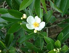
Micrococcus Yunnanesis is one of six species of genus Micrococcus. Strains of this microbe have been re-classified numerous times with previous names such as Microccoccus luteus and Sarcina subflava. It's a Gram-positive cocci that can be found isolated from plant roots of Polyspora Axillaris.
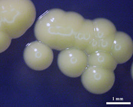
It's colony morphology is beige, smooth appearance, and circular with entire margins. Disinfectant sensitivity with small inhibition by cloves and lysol. No antimicrobial sensitivity detected.
Cellular morphology is in tetrads, irregular clusters, and pairs 
Genome Structure
Our Genome came out to: NNNNNNNNNNNNNNNNNNNNNNNNNNNNNNNNNNNNGNNGCNCGACGCCGCNNGNGTTGACGGCCNCCNNTATTGTAAACCTCTTTCNTAGGGAAGAAGCTAAATTGACGGTACCTGCNNAATAAGCACCGGCTAACTACGTGCCAACAGCCGCGGTAATACGTAGGGTGCGAGCGTTATCCGGAATTATTGGGC CTAAAGAGCTTNNATGCGGTTTGTCGCGTCTGTCGTGAAANNCCGGGGCTTAACCCCGGATCTGCGGTGGGTACGGGCACACTANACTGCAGTANGGAAGACTGGAATTCCTGGTGTAGCGCTGGAATGCCCACATATCAGGAGGAACACCGATGGCGAAGGCAGGTCTCTGGGCTGTAACTGACGCTGAGGAGC GAAAGCATGGGGAGCGAACAGGATTATATACCCTGGTAGTCCATGCCGAAAACGTTGGGCACTAGGTGTGGGGACCATTCCACGGTTTCCGCGCCGCACCTAACGCATTAAGTGCCCCGCCTGGGCAGTACNGCCGCAAGGCTACAACTCAAAGGAATTGACGGGNGCCCGCACAAGCGGCGGAGCATGCGGATT AATTCGATGCANCGCGAANAACCTTACCAAGGCTTGANNTGTTCTCNATCGCCGTANANATACCGTTTNCCCTTTNNNGCGGGNTCNCNNNTGGTGCATGGTTGTCGNCAGCTCGTGTCNTGATATCTNNTAGCTGTTTNCTNGNNNNNNTNNANNNNNNNNNNNNNNNNNNNNNNNNNNNNNNNNNNNNNNGNN GCNCGACGCCGCNNGNGTTGACGGCCNCCNNTATTGTAAACCTCTTTCNTAGGGAAGAAGCTAAATTGACGGTACCTGCNNAATAAGCACCGGCTAACTACGTGCCAACAGCCGCGGTAATACGTAGGGTGCGAGCGTTATCCGGAATTATTGGGCCTAAAGAGCTTNNATGCGGTTTGTCGCGTCTGTCGTGAA NNCCGGGGCTTAACCCCGGATCTGCGGTGGGTACGGGCACATANACTGCAGTANGGAAGACTGGAATTCCTGGTGTAGCGCTGGAATGCCCACATATCAGGAGGAACACCGATGGCGAAGGCAGGTCTCTGGGCTGTAACTGACGCTGAGGAGCGAAAGCATGGGGAGCGAACAGGATTATATACCCTGGTAGTC CATGCCGAAAACGTTGGGCACTAGGTGTGGGGACCATTCCACGGTTTCCGCGCCGCACCTAACGCATTAAGTGCCCCGCCTGGGCAGTACNGCCGCAAGGCTACAACTCAAAGGAATTGACGGGNGCCCGCACAAGCGGCGGAGCATGCGGATTAATTCGATGCANCGCGAANAACCTTACCAAGGCTTGANNTG TTCTCNATCGCCGTANANATACCGTTTNCCCTTTNNNGCGGGNTCNCNNNTGGTGCATGGTTGTCGNCAGCTCGTGTCNTGATATCTNNTAGCTGTTTNCTNGNNNNNNTNNAN. Not pretty or accurate.
M. Yunnanensis resembles other microccoci to a degree of 65.4 % in it's DNA structure. A sequencing of 16S rRNA gene was performed and it matched with M. Luteus at a 99.71% ratio. The ratios matched almost identically to other microccoci. The only notable exception to its structure was within its major fatty acids with iso-C 16 : 0 (14.25 %) a large number compared to the rest of the recognized Micrococcus species. With hybridazation experimentation and clear physiological differences indicate that the novel strain represents a separate genomic species. At this time, further testing is needed to indicate M. Yunannenensis' importance outside the realm of other Micrococci.
Cell Structure, Metabolism and Life Cycle
Cells are Gram-positive, aerobic, non-endospore-forming cocci (0.8–1 μm in diameter). Motility is not observed. Colonies on TSA are beige, smooth and circular with entire margins. No pigment is produced. Temperature, pH and NaCl tolerance ranges for growth are 4–45 °C, pH 6–8 and 0–15 % (w/v), respectively. Optimal growth occurs at 28 °C and about pH 7.0–8.0. Catalase-positive and oxidase-negative. Gelatin and starch are not hydrolysed. Negative for Methyl red, Voges–Proskauer reaction, urease and the reduction of nitrate.
Physiology and Pathogenesis
The cells are endophytic bacteria; defined as bacteria that colonizes the internal tissue of the plant while showing no external sign of infection or negative effect on their host. Considering the number of plants in the world, each having distinct types of entophytic bacteria, this can be suggested as common. Micrococcus generally an endopythic bacteria can be an opportunistic pathogen, particularly in hosts with compromised immune systems, such as HIV patients. It can be difficult to identify Micrococcus as the cause of an infection, since the organism is a normally present in skin microflora, and the genus is seldom linked to disease. In rare cases, death of immunocompromised patients has occurred from pulmonary infections caused by Micrococcus. However, no human or animal testing has been done or documented on M. Yunannensis.
Microccoci can be catabolically versatile, with the ability to utilize a wide range of unusual substrates, such as pyridine, herbicides, chlorinated biphenyls, and oil. They can also be used to detox or biodegrade environmental pollutants.
Notably, Yunnanensis produces a “restriction enzyme” used for cutting DNA in biotechnology applications.
References
Micrococcus yunnanensis sp. nov., a novel actinobacterium isolated from surface-sterilized Polyspora axillaris roots. "International Journal of systemic and Evolutionary Microbiology. 2009. Volume 59 p. 2383-2387
Zhuang W, Tay J, Maszenan A, Krumholz L, Tay S (2003). "Importance of Gram-positive naphthalene-degrading bacteria in oil-contaminated tropical marine sediments". Lett Appl Microbiol. 36 (4): 251–7.
Doddamani H, Ninnekar H (2001). "Biodegradation of carbaryl by a Micrococcus species". Curr Microbiol. 43 (1): 69–73.
https://commons.m.wikimedia.org/wiki/File:Polyspora_axillaris_3698.JPG
https://commons.m.wikimedia.org/wiki/File:M221.jpg#mw-jump-to-license
Author
Page authored by Thomas Dominguez and Orelia Daniels


