Chest Port Microbial Infections: Difference between revisions
| Line 23: | Line 23: | ||
Although port infections are not as common as other catheter infections, microbial infections are still the most significant complication resulting in port excision. About 5% of patients require port excision because of infection<ref>[https://www.ncbi.nlm.nih.gov/pmc/articles/PMC3036285/ Funaki, Brian. “Subcutaneous Chest Port Infection.” <i>Seminars in Interventional Radiology</i>, 22.3 (2005): 245–247. <i>PMC</i>.] </ref>. Infections of implanted devices most commonly result from <i>Staphylococcus aureus</i>, <i>Staphylococcus epidermidis</i>, <i>Enterococcus faecalis</i>, <i>Streptococcus vidrians</i>, <i>Klebsiella pneumonia</i>, and <i>Pseudomona aeruginosa</i>. <ref> [https://www.ncbi.nlm.nih.gov/pubmed/24515846 Paredes, J.,Alonso-Acre, M., Schmidt, C., Valderas, D., Sedano, B., Legarda, J., Arizti, F., Gomez, E., Aguinaga, A., Del Pozo, J.L., Arana, S. "Smart central venous port for early detection of bacterial biofilm related infections" <i> Biomed Microdevices</i>, 16(2014): 365.] </ref> Of the above microbes, <i>S. epidermidis</i> is the most relevant port associated pathogen. In the United States, Jukes et al. estimate 80% of nosocomial catheter related bloodstream infections (CRBSI) are a result of <i>S. epidermidis</i> <ref>[https://www.ncbi.nlm.nih.gov/pubmed/20813851 Jukes, L., Mikhail, J., Bome-Mannathoko, N., Hadfield, S.J., Harris, L.G., El-Bouri, K., Davies, A.P., Mack, D. "Rapid differentiation of <i>Staphylococcus aureus</i>, <i>Staphylococcus epidermidis</i> and other coagulase-negative staphylococci and meticillin susceptibility testing directly from growth-positive blood cultures by multiplex real-time PCR" <i>J. Med. Microbiol</i>, 59 (2010):1456–1461] </ref> . <br> | Although port infections are not as common as other catheter infections, microbial infections are still the most significant complication resulting in port excision. About 5% of patients require port excision because of infection<ref>[https://www.ncbi.nlm.nih.gov/pmc/articles/PMC3036285/ Funaki, Brian. “Subcutaneous Chest Port Infection.” <i>Seminars in Interventional Radiology</i>, 22.3 (2005): 245–247. <i>PMC</i>.] </ref>. Infections of implanted devices most commonly result from <i>Staphylococcus aureus</i>, <i>Staphylococcus epidermidis</i>, <i>Enterococcus faecalis</i>, <i>Streptococcus vidrians</i>, <i>Klebsiella pneumonia</i>, and <i>Pseudomona aeruginosa</i>. <ref> [https://www.ncbi.nlm.nih.gov/pubmed/24515846 Paredes, J.,Alonso-Acre, M., Schmidt, C., Valderas, D., Sedano, B., Legarda, J., Arizti, F., Gomez, E., Aguinaga, A., Del Pozo, J.L., Arana, S. "Smart central venous port for early detection of bacterial biofilm related infections" <i> Biomed Microdevices</i>, 16(2014): 365.] </ref> Of the above microbes, <i>S. epidermidis</i> is the most relevant port associated pathogen. In the United States, Jukes et al. estimate 80% of nosocomial catheter related bloodstream infections (CRBSI) are a result of <i>S. epidermidis</i> <ref>[https://www.ncbi.nlm.nih.gov/pubmed/20813851 Jukes, L., Mikhail, J., Bome-Mannathoko, N., Hadfield, S.J., Harris, L.G., El-Bouri, K., Davies, A.P., Mack, D. "Rapid differentiation of <i>Staphylococcus aureus</i>, <i>Staphylococcus epidermidis</i> and other coagulase-negative staphylococci and meticillin susceptibility testing directly from growth-positive blood cultures by multiplex real-time PCR" <i>J. Med. Microbiol</i>, 59 (2010):1456–1461] </ref> . <br> | ||
<i>S. epidermidis</i> is a natural member of the human skin | <i>S. epidermidis</i> is a natural member of the human skin flora<ref> [https://www.ncbi.nlm.nih.gov/pmc/articles/PMC4330918/ Buttner, H., Dietrich, M. and H. Rohde. "Structural Basis of <i>Staphylococcus Epidermidis</i> Biofilm Formation: Mechanisms and Molecular Interactions." <i>Frontiers in Cellular and Infection Microbiology</i> 5 (2015): 14. <i>PMC</i>.] </ref]. Under normal conditions, <i>S. epidermidis</i> is not pathogenic. <i>S. epidermidis</i> only acts as a human pathogen in individuals with compromised immune systems, immunosuppression, or chemotherapy related neutropenia<ref> [https://www.ncbi.nlm.nih.gov/pmc/articles/PMC4330918/ Buttner, H., Dietrich, M. and H. Rohde. "Structural Basis of <i>Staphylococcus Epidermidis</i> Biofilm Formation: Mechanisms and Molecular Interactions." <i>Frontiers in Cellular and Infection Microbiology</i> 5 (2015): 14. <i>PMC</i>.] </ref]. <br> | ||
Common port infections include <i>S. epidermidis</i> biofilm formation inside the catheter lumen2. Biofilm formation is threefold. <i> S. epidermidis</i> adhere to the catheter surface to be colonized, a microcolony forms, and <i>S. epidermidis</i> cells detach from a mature biofilm allowing <i>S. epidermidis</i> colonization on additional body sites. <br> | Common port infections include <i>S. epidermidis</i> biofilm formation inside the catheter lumen2. Biofilm formation is threefold. <i> S. epidermidis</i> adhere to the catheter surface to be colonized, a microcolony forms, and <i>S. epidermidis</i> cells detach from a mature biofilm allowing <i>S. epidermidis</i> colonization on additional body sites. <br> | ||
Revision as of 22:04, 12 April 2017
Section

By [Hannah Lorico Hertz]
At right is a sample image insertion. It works for any image uploaded anywhere to MicrobeWiki.
The insertion code consists of:
Double brackets: [[
Filename: PHIL_1181_lores.jpg
Thumbnail status: |thumb|
Pixel size: |300px|
Placement on page: |right|
Legend/credit: Electron micrograph of the Ebola Zaire virus. This was the first photo ever taken of the virus, on 10/13/1976. By Dr. F.A. Murphy, now at U.C. Davis, then at the CDC.
Closed double brackets: ]]
Other examples:
Bold
Italic
Subscript: H2O
Superscript: Fe3+
Introduce the topic of your paper. What is your research question? What experiments have addressed your question? Applications for medicine and/or environment?
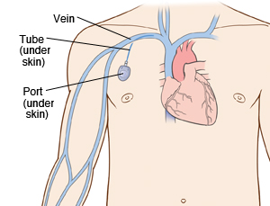
A chest port is a catheter connected to a reservoir inserted under the skin of the chest and used to administer medicines directly into a vein over a long period of time. Chest ports are commonly used to administer long-term chemotherapy in children because of the ease of care for port maintenance. In comparison to an IV line, chest ports can stay in place for months at a time, can be used to collect blood samples without needles, and have a lower risk of infection over time.
Although port infections are not as common as other catheter infections, microbial infections are still the most significant complication resulting in port excision. About 5% of patients require port excision because of infection[1]. Infections of implanted devices most commonly result from Staphylococcus aureus, Staphylococcus epidermidis, Enterococcus faecalis, Streptococcus vidrians, Klebsiella pneumonia, and Pseudomona aeruginosa. [2] Of the above microbes, S. epidermidis is the most relevant port associated pathogen. In the United States, Jukes et al. estimate 80% of nosocomial catheter related bloodstream infections (CRBSI) are a result of S. epidermidis [3] .
S. epidermidis is a natural member of the human skin floraCite error: Closing </ref> missing for <ref> tag
[4]
A citation code consists of a hyperlinked reference within "ref" begin and end codes.
Section 1
Include some current research, with at least one figure showing data.
Every point of information REQUIRES CITATION using the citation tool shown above.
Section 2
Include some current research, with at least one figure showing data.
Section 3
Include some current research, with at least one figure showing data.
Section 4
Conclusion
References
- ↑ Funaki, Brian. “Subcutaneous Chest Port Infection.” Seminars in Interventional Radiology, 22.3 (2005): 245–247. PMC.
- ↑ Paredes, J.,Alonso-Acre, M., Schmidt, C., Valderas, D., Sedano, B., Legarda, J., Arizti, F., Gomez, E., Aguinaga, A., Del Pozo, J.L., Arana, S. "Smart central venous port for early detection of bacterial biofilm related infections" Biomed Microdevices, 16(2014): 365.
- ↑ Jukes, L., Mikhail, J., Bome-Mannathoko, N., Hadfield, S.J., Harris, L.G., El-Bouri, K., Davies, A.P., Mack, D. "Rapid differentiation of Staphylococcus aureus, Staphylococcus epidermidis and other coagulase-negative staphylococci and meticillin susceptibility testing directly from growth-positive blood cultures by multiplex real-time PCR" J. Med. Microbiol, 59 (2010):1456–1461
- ↑ Bartlett et al.: Oncolytic viruses as therapeutic cancer vaccines. Molecular Cancer 2013 12:103.
Authored for BIOL 238 Microbiology, taught by Joan Slonczewski, 2017, Kenyon College.
