Nocardia farcinica: Difference between revisions
| Line 61: | Line 61: | ||
<i>Nocardia</i> are parasitic bacteria which grow and reproduce on organic material. Their man habitat is carbon-rich sources such as soils, and plant and animal tissues. In fact, they can be found almost anywhere. One environmental survey found <i>Nocardia</i> in "beach sand, swimming pools, house dust, and garden soil".(14) Upon infection of a plant or animal host, it metabolizes necrotizing tissues for energy and nutrients. Because <i>Nocardia</i> can form endospores, transmission of the bacteria "aerogenically"(2) from one host to another is relatively easy, and the bacteria can survive dormantly when food sources are not present.<br> | <i>Nocardia</i> are parasitic bacteria which grow and reproduce on organic material. Their man habitat is carbon-rich sources such as soils, and plant and animal tissues. In fact, they can be found almost anywhere. One environmental survey found <i>Nocardia</i> in "beach sand, swimming pools, house dust, and garden soil".(14) Upon infection of a plant or animal host, it metabolizes necrotizing tissues for energy and nutrients. Because <i>Nocardia</i> can form endospores, transmission of the bacteria "aerogenically"(2) from one host to another is relatively easy, and the bacteria can survive dormantly when food sources are not present.<br> | ||
<br> | <br> | ||
As stated, <i>Nocardia</i> infections can be transmitted aerogenically to host respiratory systems or cutaneous wound sites, they can be introduced by innoculation (puncture wounds with the bacteria contaminant), and through fluid contact (ex: case reported of keratitis due to contaminated contact lense).(12) Infection, once contracted, may spread systemically (dissemination). Disseminated infections often are the result of pulmonary infections, and are the most life threatening. (14) <i>N. farcinica</i> is | As stated, <i>Nocardia</i> infections can be transmitted aerogenically to host respiratory systems or cutaneous wound sites, they can be introduced by innoculation (puncture wounds with the bacteria contaminant), and through fluid contact (ex: case reported of keratitis due to contaminated contact lense).(12) Infection, once contracted, may spread systemically (dissemination). Disseminated infections often are the result of pulmonary infections, and are the most life threatening. (14) <i>N. farcinica</i> is an opportunistic, yet virulent in general, and shown to be the most virulent of <i>Nocardia</i> species studied. (13)<br> | ||
<br> | <br> | ||
<i>Nocardia</i> infections have a high mortality rate, are resistant to treatement, and are particularly persistent and harmful in patients who have a suppressed immune system (as with HIV or transplant patients).(14) Drugs that show some degree of effect against <i>Nocardia</i> include ketoconazole, clotrimazole, and miconazole. (3)<br> | <i>Nocardia</i> infections have a high mortality rate, are resistant to treatement, and are particularly persistent and harmful in patients who have a suppressed immune system (as with HIV or transplant patients).(14) Drugs that show some degree of effect against <i>Nocardia</i> include ketoconazole, clotrimazole, and miconazole. (3)<br> | ||
Revision as of 06:41, 5 June 2007
A Microbial Biorealm page on the genus Nocardia farcinica
Classification
Higher order taxa
Superkingdom: Bacteria
Phylum: Actinobacteria
Class: Actinobacteria
Subclass: Actinobacteridae
Order: Actinomycetales
Suborder: Corynebacterineae
Family: Nocardiaceae
Genus: Nocardia
(1)
Species
Nocardia farcinica
(1)
Description and significance
Species of the Nocardia Genus are Gram positive bacteria. Their cell envelope consists of a peptidoglycan cell wall, inside of which lay the cell membrane; a lipid bilayer with imbedded proteins. They are saprophytic; their habitats vary based on their need to grow and live on dead and/or decaying organic material. Soils, plants, animal tissues, and human tissues make ideal environments for growth. (3)
Farcinica are rod shaped but can fragment into coccoids. They are filamentous, produce endospores, are non motile, are obligate aerobes, and experience optimal growth at body temperature. (3,4) Nocardia colonies vary from white, to tan, orange and red in color. An image of Nocardia farcinica is not available for publication on MicrobeWiki at the date of this entry, but the following are provided for comparison:
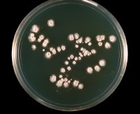
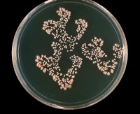
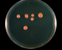
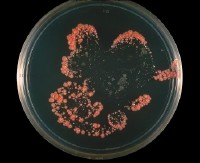
All four images above show Nocardia asteroides grown on the same media, at the same temperature. Variances in size, color, and shape of colonies are apparent.
Images provided by CDC/Dr. William Kaplan
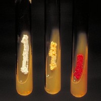
Left tube: Actinomadura madurae, Middle tube: Nocardia asteroides, Right tube: Micromonspora.
Image provided by CDC/Dr. David Berd
Nocardia farcinica images that are not available for publication on this site show colonies that are golden, to light, medium, and dark orange. The colonies appear globular and shiny with surfaces ranging from smooth to reticulated. (5,6) [1] and [2]
The genus Nocardia was first discovered Edmund Nocard, a French veterinarian, in 1889 as the cause of bovine infections. Nocardia farcinica is significant to modern medicine as it is a pathogen which threatens both human health and the health of important assets such as herds of cattle, horses, and many other animals. It is particularly resistant to treatment, and a high mortality rate among those infected. (7)
Genome structure
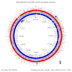
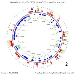
The genome of Nocardia farcinica containes a circular chromosome that is 6,021,225 base pairs long comprising 5,811 genes (8), and has a GC content of about 71%. Farcinica also has two plasmids; pNF1 & pNF2. pNF1 is 184,027 base pairs long, comprising 160 genes (8), and has a GC content of about 67%, while pNF2 is 87,093 base pairs long, comprising 93 genes(8), and its GC content is about 68%. (3) The defining characteristics about the various Nocardia species, including farcinica have been determined mainly by genomic sequencing of the 16S rRNA. (9)
Below is BacMap Genome Mapping, courtesy of Stothard P, Van Domselaar G, Shrivastava S, Guo A, O'Neill B, Cruz J, Ellison M, Wishart DS (2005) BacMap: an interactive picture atlas of annotated bacterial genomes. Nucleic Acids Res 33:D317-D320.
Detailed genomic information is available through BacMap [3]
 ****
**** ****
****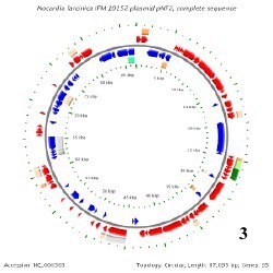
Image 1: N. farcinica Chromosome
Image 2: pNF1 (Plasmid 1)
Image 3: pNF2 (Plasmid 2)
Cell structure and metabolism
Basic cell structure of N. farcinica:
Rod shaped, Gram positive, Mycolic acid cell wall, lipid bilayer cell membrane with associated proteins. The cell has one circular chromosome, and two plasmids. My research shows no indication of flagella, nor pilli on the N. farcinica species.
Nocardia species "form filamentous, branched cells which fragment into pleomorphic, rod-shaped, or coccoid elements... .filamentous cells protrude into the air away from the surface (aerial hyphae)"(11). All Mycolic acids comprising the cell wall of this bacteria are unsaturated, as opposed to some other Nocardia species such as asteroides who have a large quantity of saturated Mycolic acids. (10)
Metabolism:
N. farcinica carries out a broad variety of metabolic pathways. It's carbohydrate metabolism includes glycolysis, TCA cycle, pentose phosphate, and a long list of sugar metabolic pathways. Energy metabolic pathways include oxidative phosphorylation, methane, nitrogen, and sulfur metabolisms. Interestingly, N. farcinica carries out two photosynthetic pathways; the carbon fixation and reductive carboxylate cycle. (12) The research done on the metabolic pathways for N. farcinica is extensive and they can be viewed in detail on the N. farcinica site of the online Kyoto Encyclopedia of Genes and Genomes (KEGG) [4]
Secondary Metabolism:
Ishikawa, et al. describes the high quantity of oxegenases found in N. farcinica and proposes that their purposes may be for fatty acid metabolism and defenses against toxins. It was also found that N. farcinica produces "bioactive molecules" through both the "mevalonate and the 'non' mevalonate pathways". (3)
Ecology & Pathology
Nocardia are parasitic bacteria which grow and reproduce on organic material. Their man habitat is carbon-rich sources such as soils, and plant and animal tissues. In fact, they can be found almost anywhere. One environmental survey found Nocardia in "beach sand, swimming pools, house dust, and garden soil".(14) Upon infection of a plant or animal host, it metabolizes necrotizing tissues for energy and nutrients. Because Nocardia can form endospores, transmission of the bacteria "aerogenically"(2) from one host to another is relatively easy, and the bacteria can survive dormantly when food sources are not present.
As stated, Nocardia infections can be transmitted aerogenically to host respiratory systems or cutaneous wound sites, they can be introduced by innoculation (puncture wounds with the bacteria contaminant), and through fluid contact (ex: case reported of keratitis due to contaminated contact lense).(12) Infection, once contracted, may spread systemically (dissemination). Disseminated infections often are the result of pulmonary infections, and are the most life threatening. (14) N. farcinica is an opportunistic, yet virulent in general, and shown to be the most virulent of Nocardia species studied. (13)
Nocardia infections have a high mortality rate, are resistant to treatement, and are particularly persistent and harmful in patients who have a suppressed immune system (as with HIV or transplant patients).(14) Drugs that show some degree of effect against Nocardia include ketoconazole, clotrimazole, and miconazole. (3)
Patient symptoms:
Pulmonary Nocardiosis:
-Cough with sputum, increasing difficulty breathing, loss of weight, chest and joint pain, night sweats, liver and spleen enlargement, general malaise, and headache and skin rashes/lumps.
Brain Nocardiosis:
-Fever, headache, loss of neurological function, sometimes asymptomatic.
Cutaneous Nocardiosis:
-Ulcers/Nodules of infection which may follow lymph node chains, progressive towards muscle and bone.
(15)
Occular Nocardiosis:
-Keratitis.
(12)
Application to Biotechnology
My research indicates that all that has been suggested regarding the species' usefulness in biotechnology is that some of its genes "are possibly involved in the production of novel peptides with biological activity, such as thiazolyl peptide antibiotics previously isolated from Nocardia sp."(3)
Being that this species is a very virulent pathogen, and was sequenced only three years ago, it seems logical that there has not yet been any clear evidence of biological benefit. As research continues, this may change.
Current Research
(Sample of some current research as of June 5th, 2007)
"The complete genomic sequence of Nocardia farcinica IFM 10152" -2004
Summary - The researchers determined the genomic sequence of the single circular chromosome of N. farcinica along with its two plasmids in 2004. Genese sequenced give valuable starting points in determining virulence, drug susceptibilities, and to further research the species' secondary metabolism. Three drugs were found to have an affect on N. farcinica; clotrimazole, ketoconazole, and miconazole. Data suggested that the bacterium's sterol pathway may be a target for drug development.(3)
"Clinical and laboratory features of the Nocardia spp. based on current molecular taxonomy." -2006
Summary - Due to ongoing discovery of distinct species within the Nocardia genus, a great amount of the current research being done has to do with genetic sequencing. Genomic sequencing is performed using the 16S rRNA sequencing method. Sequencing of new species is of importance because of the virulence of the Nocardia genus, and thus the need to develop drug treatments to Nocardia infections. Accurate and fast methods of distinguishing between the various species are being researched in order to most efficiently treat patients. This article outlines what is currently known of the virulent Nocardia species, describes what treatments are effective, and also means to distinguish between the Nocardia species.(9)
"Analysis of secA1 Gene Sequences for Identification of Nocardia Species" -2006
Summary - This article describes an alternative way of distinguishing between Nocardia species, other than the 16S rRNA method. This alternative way is by analyzing a portion of the secA1 gene. Sequence analysis of this gene showed distinct base pairing between all 30 species tested, unique to each species. It was also said that the 16S rRNA method was not quite as distinct, as some species showed similarities to others. Researchers suggest that this new, alternate method, will prove to be more accurate.(16)
References
1. Wheeler DL, Chappey C, Lash AE, Leipe DD, Madden TL, Schuler GD, Tatusova TA, Rapp BA (2000). Database resources of the National Center for Biotechnology Information. Nucleic Acids Res 2000 Jan 1;28(1):10-4 [5]
2. J. Blümel, E. Blümel, A. F. Yassin, H. Schmidt-Rotte, and K. P. Schaal1. Typing of Nocardia farcinica by Pulsed-Field Gel Electophoresis Reveals an Endemic Strain as Source of Hospital Infections. J Clin Microbiol. 1998 January; 36(1): 118–122. [6]
3. Jun Ishikawa, Atsushi Yamashita, Yuzuru Mikami, Yasutaka Hoshino, Haruyo Kurita, Kunimoto Hotta, Tadayoshi Shiba, and Masahira Hattori. The complete genomic sequence of Nocardia farcinica IFM 10152. 2004 Oct 10.1073/pnas.0406410101. [7]
4. NCBI Genome Project > Nocardia farcinica IFM 10152 [NCBI]
5. Dept. of Bioactive Molecules, Nat Inst of Infectious Disease. Nocardia farcinica Genome Project 13117. [8]
6. Digital Atlas – Atlas of Actinomycetes, pg 2-8. Nat Inst of Infectious Disease. [9]
7. Munoz J, Mirelis B, et al., Clinical and microbiological features of nocardiosis 1997-2003. [10]
8. Stothard P, Van Domselaar G, Shrivastava S, Guo A, O'Neill B, Cruz J, Ellison M, Wishart DS (2005) BacMap: an interactive picture atlas of annotated bacterial genomes. Nucleic Acids Res 33:D317-D320 [11]
9. Barbara A. Brown-Elliott, June M. Brown, Patricia S. Conville, and Richard J. Wallace, Jr. Clinical and Laboratory Features of the Nocardia spp. Based on Current Molecular Taxonomy. Clin Microbiol Rev. 2006 April; 19(2): 259–282. [12]
10. Nischiuchi Y, Baba T, Hotta HH, Yano I. Mycolic acid analysis in Nocardia species. The mycolic acid compositions of Nocardia asteroides, N. farcinica, and N. nova. J Microbiol Methods, 1999 Aug, 37(2):111-22. [13]
11. B L Beaman and L Beaman, Nocardia species: host-parasite relationships. Clin Microbiol Rev. 1994 April; 7(2): 213–264. [14]
12. Catharina A. Eggink, Pieter Wesseling, et al. Severe Keratitis Due to Nocardia farcinica. J of Clinical Microbiology, Apr 1997, p 99-1001. [15]
13. Edward P. Desmond, Martha Flores. Mouse pathogenicity studies of N. asteroides complex species and clinical correlation with human isolates. Volume 110 Issue 3 Page 281Issue 3 - 284 - July 1993. [16]
14. M M McNeil and J M Brown. The medically important aerobic actinomycetes: epidemiology and microbiology. Clin Microbiol Rev. 1994 July; 7(3): 357–417. [17]
15. AllRefer.com Health & A.D.A.M. Inc. [18]
16. Patricia S. Conville, Adrian M. Zelazny, and Frank G. Witebsky. Analysis of secA1 Gene SEquences for Identification of Nocardia Species. J Clin Microbiol. 2006 August; 44(8): 2760–2766. [19]
Edited by Julie Crownover, student of Rachel Larsen and Kit Pogliano
