The Structure and Function of Ebola Treatment Units: Difference between revisions
| Line 2: | Line 2: | ||
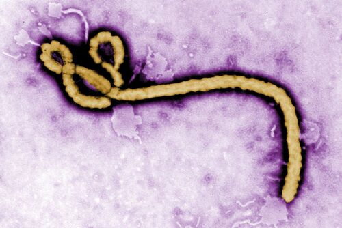
[[Image:Ebolapurpleimage.jpg|thumb|500px|right|A transmission electron microscopy image of the Ebola virion. Photo credit: [https://www.wsj.com/articles/ebola-in-congo-starts-scramble-to-contain-outbreak-1494858534/ The Wall Street Journal]]] | [[Image:Ebolapurpleimage.jpg|thumb|500px|right|A transmission electron microscopy image of the Ebola virion. Photo credit: [https://www.wsj.com/articles/ebola-in-congo-starts-scramble-to-contain-outbreak-1494858534/ The Wall Street Journal]]] | ||
The ebola virus genus which is responsible for the cause of Ebola Virus Disease (EVD), is a hemorrhagic fever virus in humans.<ref>Kadanali A, Karagoz G. An overview of Ebola virus disease. North Clin Istanb. 2015 Apr 24;2(1):81-86. doi: 10.14744/nci.2015.97269. PMID: 28058346; PMCID: PMC5175058.</ref> The U.S. Centers for Disease Control and Prevention (CDC) classifies Ebola as a Category A select agent, making it a “high-priority agent” as it threatens national security. <ref>Centers for Disease Control and Prevention. (2018a, April 4). CDC. Centers for Disease Control and Prevention. https://emergency.cdc.gov/agent/agentlist-category.asp</ref> Ebolaviruses (EBOV) belong to the virus family Filoviridae which it shares with Marburgvirus and Cuevavirus.<ref>Kadanali A, Karagoz G. An overview of Ebola virus disease. North Clin Istanb. 2015 Apr 24;2(1):81-86. doi: 10.14744/nci.2015.97269. PMID: 28058346; PMCID: PMC5175058.</ref> EBOV genomes consist of non-segmented, single-stranded RNA structures that includes seven open reading frames.<ref>Postigo-Hidalgo, I., Fischer, C., Moreira-Soto, A., Tscheak, P., Nagel, M., Eickmann, M., & Drexler, J. F. (2020, January). Pre-emptive genomic surveillance of emerging ebolaviruses. Euro surveillance : bulletin Europeen sur les maladies transmissibles = European communicable disease bulletin. https://www.ncbi.nlm.nih.gov/pmc/articles/PMC6988270/<.ref>[15] The EBOV genome is approximately 18.9 kilobases long.[5] | The ebola virus genus which is responsible for the cause of Ebola Virus Disease (EVD), is a hemorrhagic fever virus in humans.<ref>Kadanali A, Karagoz G. An overview of Ebola virus disease. North Clin Istanb. 2015 Apr 24;2(1):81-86. doi: 10.14744/nci.2015.97269. PMID: 28058346; PMCID: PMC5175058.</ref> The U.S. Centers for Disease Control and Prevention (CDC) classifies Ebola as a Category A select agent, making it a “high-priority agent” as it threatens national security. <ref>Centers for Disease Control and Prevention. (2018a, April 4). CDC. Centers for Disease Control and Prevention. https://emergency.cdc.gov/agent/agentlist-category.asp</ref> Ebolaviruses (EBOV) belong to the virus family Filoviridae which it shares with Marburgvirus and Cuevavirus.<ref>Kadanali A, Karagoz G. An overview of Ebola virus disease. North Clin Istanb. 2015 Apr 24;2(1):81-86. doi: 10.14744/nci.2015.97269. PMID: 28058346; PMCID: PMC5175058.</ref> EBOV genomes consist of non-segmented, single-stranded RNA structures that includes seven open reading frames.<ref>Postigo-Hidalgo, I., Fischer, C., Moreira-Soto, A., Tscheak, P., Nagel, M., Eickmann, M., & Drexler, J. F. (2020, January). Pre-emptive genomic surveillance of emerging ebolaviruses. Euro surveillance : bulletin Europeen sur les maladies transmissibles = European communicable disease bulletin. https://www.ncbi.nlm.nih.gov/pmc/articles/PMC6988270/ <.ref>[15] The EBOV genome is approximately 18.9 kilobases long.[5] | ||
The EBOV genome encodes for seven genes each containing its own open reading frame.[3][6][28] The seven gene all aid in the viral entry and replication of the virus (Figure ): | The EBOV genome encodes for seven genes each containing its own open reading frame.[3][6][28] The seven gene all aid in the viral entry and replication of the virus (Figure ): | ||
Glycoproteins(GPs) bind to the host's receptors on the cell's surface to insert its viral genome. | Glycoproteins(GPs) bind to the host's receptors on the cell's surface to insert its viral genome. | ||
Revision as of 19:26, 14 April 2024
Introduction

The ebola virus genus which is responsible for the cause of Ebola Virus Disease (EVD), is a hemorrhagic fever virus in humans.[1] The U.S. Centers for Disease Control and Prevention (CDC) classifies Ebola as a Category A select agent, making it a “high-priority agent” as it threatens national security. [2] Ebolaviruses (EBOV) belong to the virus family Filoviridae which it shares with Marburgvirus and Cuevavirus.[3] EBOV genomes consist of non-segmented, single-stranded RNA structures that includes seven open reading frames.<ref>Postigo-Hidalgo, I., Fischer, C., Moreira-Soto, A., Tscheak, P., Nagel, M., Eickmann, M., & Drexler, J. F. (2020, January). Pre-emptive genomic surveillance of emerging ebolaviruses. Euro surveillance : bulletin Europeen sur les maladies transmissibles = European communicable disease bulletin. https://www.ncbi.nlm.nih.gov/pmc/articles/PMC6988270/ <.ref>[15] The EBOV genome is approximately 18.9 kilobases long.[5] The EBOV genome encodes for seven genes each containing its own open reading frame.[3][6][28] The seven gene all aid in the viral entry and replication of the virus (Figure ): Glycoproteins(GPs) bind to the host's receptors on the cell's surface to insert its viral genome. The Ebola Matrix Protein (VP40 NPs) gives EBOV its filamentous shape and directs the budding of enveloped viruses Nucleoproteins are subunits of the Nucleocapsid that as whole protects the viral genome. VP35, VP30, and VP24 proteins play roles in the formation of EBOVs helical nucleocapsid The Polymerase (L) Protein is responsible for creating several copies of the RNA genome within hosts cells
GPs have been identified as a target for vaccine development, as their outward position on EBOV surfaces make them visible to antibodies.[3] A major hope of vaccine development for EVD is to create a treatment that allows the binding of antibodies to non-carbohydrated regions of viral protein.[3] As of 2019, the very first FDA-approved Ebola vaccine called Ervebo has been approved and released for the use of people eighteen years or older.[25] Exposure to the GP in humans allows for the neutralization of antibodies, along with the restraint of viral attachment and fusion to host receptors.[24][26] Although Ervebo has saved many lives, the vaccine merely promotes the immunity of Zaire ebolavirus[24] With the possibility of high mutation rate frequencies of the EBOV genome and other infectious species, vaccine development is still underway in pharmaceutical companies.[27]
Symptoms
The onset of symptoms typically occurs between the incubation period of 2-21 days after exposure to EBOV.[9] At its early to mild stages, victims may experience the following flu-like symptoms: Vomiting, fever, headache, abdominal pain and diarrhea. Advanced EVD is indicative of the following symptoms all of which are fatal, bleeding from all orifices of the body, multi-organ failure, hypovolemic shock and coffee ground emesis.[10]
Transmission
- ↑ Kadanali A, Karagoz G. An overview of Ebola virus disease. North Clin Istanb. 2015 Apr 24;2(1):81-86. doi: 10.14744/nci.2015.97269. PMID: 28058346; PMCID: PMC5175058.
- ↑ Centers for Disease Control and Prevention. (2018a, April 4). CDC. Centers for Disease Control and Prevention. https://emergency.cdc.gov/agent/agentlist-category.asp
- ↑ Kadanali A, Karagoz G. An overview of Ebola virus disease. North Clin Istanb. 2015 Apr 24;2(1):81-86. doi: 10.14744/nci.2015.97269. PMID: 28058346; PMCID: PMC5175058.
