Ophiocordyceps Fungal Invasion of Ants: Difference between revisions
Tag: Manual revert |
|||
| Line 2: | Line 2: | ||
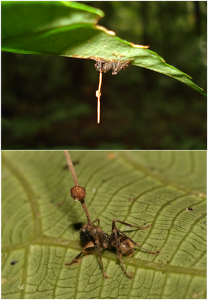
Parasitic ant-fungal relationships have been present since the Eocene epoch 45 million years ago5. These relationships, while similar in nature and spanning the order Hymenoptera, are the result of a coevolution between parasite and host resulting in highly specific mechanisms of action. The genus Ophiocordyceps falls into this category of entomopathogens, fungi that parasitize insects. While each individual species of Ophiocordyceps infects a different host ant species, the method of fungal reproduction and spreading of spores remains the same. Fungal spores penetrate the protected cuticle of the ant and begin to reproduce within internal ant tissues. After a certain period of time, usually after a few days, the infected ant will leave the colony, abandon its tasks, climb a vegetative structure, and bite on to the leaf, stem, or branch with its mandibles. Here, the ant will die, remaining latched on, as the fungus completes its life cycle by growing a fruiting body from the ant cadaver to disperse spores.<br> | Parasitic ant-fungal relationships have been present since the Eocene epoch 45 million years ago5. These relationships, while similar in nature and spanning the order Hymenoptera, are the result of a coevolution between parasite and host resulting in highly specific mechanisms of action. The genus Ophiocordyceps falls into this category of entomopathogens, fungi that parasitize insects. While each individual species of Ophiocordyceps infects a different host ant species, the method of fungal reproduction and spreading of spores remains the same. Fungal spores penetrate the protected cuticle of the ant and begin to reproduce within internal ant tissues. After a certain period of time, usually after a few days, the infected ant will leave the colony, abandon its tasks, climb a vegetative structure, and bite on to the leaf, stem, or branch with its mandibles. Here, the ant will die, remaining latched on, as the fungus completes its life cycle by growing a fruiting body from the ant cadaver to disperse spores.<br> | ||
<br><br> | <br><br> | ||
[[ | [[File:Pone.0004835.g001.PNG L.png|thumb|300px|left|Figure 1. Electron micrograph of the Ebola Zaire virus. This was the first photo ever taken of the virus, on 10/13/1976. By Dr. F.A. Murphy, now at U.C. Davis, then at the CDC.[https://phil.cdc.gov/details.aspx?pid=1833].]] | ||
<br>At right is a sample image insertion. It works for any image uploaded anywhere to MicrobeWiki. The insertion code consists of: | <br>At right is a sample image insertion. It works for any image uploaded anywhere to MicrobeWiki. The insertion code consists of: | ||
Revision as of 16:56, 11 December 2024
Introduction
Parasitic ant-fungal relationships have been present since the Eocene epoch 45 million years ago5. These relationships, while similar in nature and spanning the order Hymenoptera, are the result of a coevolution between parasite and host resulting in highly specific mechanisms of action. The genus Ophiocordyceps falls into this category of entomopathogens, fungi that parasitize insects. While each individual species of Ophiocordyceps infects a different host ant species, the method of fungal reproduction and spreading of spores remains the same. Fungal spores penetrate the protected cuticle of the ant and begin to reproduce within internal ant tissues. After a certain period of time, usually after a few days, the infected ant will leave the colony, abandon its tasks, climb a vegetative structure, and bite on to the leaf, stem, or branch with its mandibles. Here, the ant will die, remaining latched on, as the fungus completes its life cycle by growing a fruiting body from the ant cadaver to disperse spores.

At right is a sample image insertion. It works for any image uploaded anywhere to MicrobeWiki. The insertion code consists of:
Double brackets: [[
Filename: PHIL_1181_lores.jpg
Thumbnail status: |thumb|
Pixel size: |300px|
Placement on page: |right|
Legend/credit: Electron micrograph of the Ebola Zaire virus. This was the first photo ever taken of the virus, on 10/13/1976. By Dr. F.A. Murphy, now at U.C. Davis, then at the CDC.
Closed double brackets: ]]
Other examples:
Bold
Italic
Subscript: H2O
Superscript: Fe3+
Section 1 Genetics
Section titles are optional.
[1]
Include some current research, with at least one image.
Call out each figure by number (Fig. 1).
Sample citations: [1]
[2]
A citation code consists of a hyperlinked reference within "ref" begin and end codes.
For multiple use of the same inline citation or footnote, you can use the named references feature, choosing a name to identify the inline citation, and typing [4]
Second citation of Ref 1: [1]
Here we cite April Murphy's paper on microbiomes of the Kokosing river. [5]
Section 2 Microbiome
Include some current research, with a second image.
Here we cite Murphy's microbiome research again.[5]
Conclusion
You may have a short concluding section.
Overall, cite at least 5 references under References section.
References
- ↑ 1.0 1.1 1.2 Kurokawa C, Lynn GE, Pedra JH, Pal U, Narasimhan S, Fikrig E. Interactions between Borrelia burgdorferi and ticks. Nature Reviews Microbiology. 2020 Oct;18(10):587-600.
- ↑ Bartlett et al.: Oncolytic viruses as therapeutic cancer vaccines. Molecular Cancer 2013 12:103.
- ↑ Lee G, Low RI, Amsterdam EA, Demaria AN, Huber PW, Mason DT. Hemodynamic effects of morphine and nalbuphine in acute myocardial infarction. Clinical Pharmacology & Therapeutics. 1981 May;29(5):576-81.
- ↑ 4.0 4.1 text of the citation
- ↑ 5.0 5.1 Murphy A, Barich D, Fennessy MS, Slonczewski JL. An Ohio State Scenic River Shows Elevated Antibiotic Resistance Genes, Including Acinetobacter Tetracycline and Macrolide Resistance, Downstream of Wastewater Treatment Plant Effluent. Microbiology Spectrum. 2021 Sep 1;9(2):e00941-21.
Edited by [Author Name], student of Joan Slonczewski for BIOL 116, 2024, Kenyon College.
