Entamoeba histolytica: Difference between revisions
mNo edit summary |
No edit summary |
||
| (18 intermediate revisions by 2 users not shown) | |||
| Line 1: | Line 1: | ||
{{Uncurated}} | |||
{{Biorealm Genus}} | {{Biorealm Genus}} | ||
| Line 5: | Line 6: | ||
===Higher order taxa=== | ===Higher order taxa=== | ||
Cellular organisms; Eukaryota; Entamoebidae; Entamoeba | |||
===Species=== | ===Species=== | ||
''Entamoeba histolytica'' | ''Entamoeba histolytica'' | ||
{| | |||
| height="10" bgcolor="#FFDF95" | | |||
'''NCBI: [http://www.ncbi.nlm.nih.gov/Taxonomy/Browser/wwwtax.cgi?mode=Info&id=294381&lvl=3&lin=f&keep=1&srchmode=1&unlock] Taxonomy''' | |||
|} | |||
==Description and Significance== | |||
''Entamoeba histolytica'' is an anaerobic parasitic protozoan that infects the digestive tract of predominantly humans and other primates. It is a parasite that infects an estimated 50 million people around the world and is a significant cause of morbidity and mortality in developing countries. Analysis of the genome allows new insight into the workings and genome evolution of a human pathogen. | |||
Transmission of the parasite occurs when a person ingests food/water that has been contaminated with infected feces. The infection ''E. histolytica'' is called Amebiasis (or Amoebiasis). Cysts of the parasite are the viable form outside the host. They can survive weeks in water, soils and on foods under moist conditions. Once inside the host, cysts divide into four trophozoites in the small intestines. ''E. histolytica'' strains have been isolated from foreigners outside the U.S., homosexual men, patients with HIV and cynomolgus monkeys. | |||
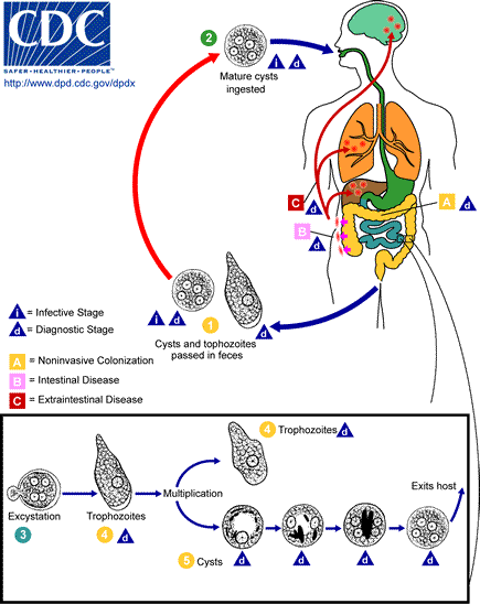
[[Image:Ambiasis_LifeCycle.gif|frame|Image of life cycle of E. histolytica parasite. Courtesy of http://www.dpd.cdc.gov/dpdx/html/Amebiasis.htm ]] | |||
==Genome structure== | |||
''E. histolytica'' (strain HM-1:IMSS) was the first human amoeba to have its genome sequenced, and analyzed. The sequencing project was a collaborative effort between TIGR and The Sanger Institute. With the difficulties in condensing and resolving the chromosomes of ''E histolytica'' by pulse-field gel electrophoresis, the exact number of chromosomes in is unknown. | |||
49% of the genome is comprised of approximately 9,938 predicted genes with an average size of 1.17 kb. One third of this organism’s predicted proteins (31.8%) do not have identifiable sequence homologues in other species. The haploid genome of strain HM-1 is less than 20 Mb in 14 chromosomes. | |||
==Cell structure and metabolism== | |||
The cysts of ''E. histolytica'' measure between 10 to 20 m in diameter. The telltale feature of the cyst is the number of nuclei; cysts will have up to four nuclei. Also, the peripheral chromatin is generally evenly distributed. | |||
The trophozoites of ''E. histolytica'' have a diameter of 15 to 20 m. They are spherical or oval shaped with a thin cell membrane and a single nucleus with a prominent nuclear border, central karysome and vacuole. They move via finger-like pseudopods towards colon. Trophozoites ingest red blood cells and absorbs their nutrients to survive. Some trophozoites transform into cysts. | |||
==Ecology== | |||
Cysts are viable in water, soils and on foods. They are formed by trophozoites and excreted in feces. There they survive for weeks in moist conditions. | |||
Trophozoite contact with human cells induces a rapid influx of calcium into the contacted cell. This stops all membrane movement. The internal organization is disrupted, organelles lyse and the cell dies. The amoeba may ingest the dead cell or intake nutrients from the cell. Presence of trophozoites containing red blood cells is indicative of tissue invasion by virulent ''E. histolytica'' parasites. Ulcers created by trophozoites have a broad base that is composed of fibrin and cellular debris. Trophozoites are found on the surface of ulcers, in the exudates and in the crater. Frequently, they are found in the submucosa, muscularis propria, serosa and small veins of the submucosa. There is little inflammatory response in early ulcers, but as the ulcer widens there is an accumulation of neutrophils, lymphocytes, histiocytes, plasma cells and sometimes eosinophils. Examination of tissues of an ameboma reveal granulation tissue, fibrosis, chronic inflammatory cells and clusters of trophozoites usually concentrated in the submucosa. Occasionally (5%-10%) trophozoites penetrate the muscle and extraintestinal ambiasis can occur. Parasites penetrate portal vessels and embolize to the liver and form liver abscesses. | |||
==Pathology== | |||
The infection of ''E. histolytica'' causes the disease, Amebiasis (or Amoebiasis). ''E. histolytica'' infects the digestive tracts of predominantly humans and other primates. ''E. histolytica'' can infect dogs and cats, but these animals do not contribute significantly to transmission since they usually do not produce cysts. Cysts do not invade tissue and are shed with the host's feces. The thick protective walls allow the cysts to remain viable for several weeks in the external environment and the internal acid content of the stomach. | |||
After a viable cyst is ingested, it travels to the small intestine where excystation occurs and it divides into four trophozoites, which is the active stage of the parasite that only survives in the host and in fresh feces. These trophozoites then mature into adult trophozoites and colonize the large intestine (particularly the caecum). | |||
Contact with human cells induces a rapid influx of calcium into the contacted cell. This stops all membrane movement. The internal organization is disrupted, organelles lyse and the cell dies. The amoeba may ingest the dead cell or intake nutrients from the cell. Presence of trophozoites containing red blood cells is indicative of tissue invasion by virulent ''E. histolytica'' parasites. Ulcers created by trophozoites have a broad base that is composed of fibrin and cellular debris. Trophozoites are found on the surface of ulcers, in the exudates and in the crater. There is little inflammatory response in early ulcers, but as the ulcer widens there is an accumulation of neutrophils, lymphocytes, histiocytes, plasma cells and sometimes eosinophils. Occasionally (5%-10%) trophozoites penetrate the muscle and serous layers which perforates the intestine. Extraintestinal ambiasis can occur. Parasites penetrate portal vessels and embolize to the liver and form liver abscesses. The abscess cavity is sometimes filled with a pasty chocolate colored material. Trophozoites form new cysts which are then excreted in the stool. | |||
The symptoms of the disease is often mild, causing diarrhea and abdominal pain. Amebic dysentery—a more severe symptom can occur. Symptoms of amebic dysentery include severe stomach pain, blood and mucus in feces and high temperature fever. Seldom does the infection invade the liver and cause an abscess. | |||
==Current Research== | |||
The genome of ''E. histolytica'' is revealed and a variety of metabolic adaptations shared with two other amitochondrial protest pathogens: Giardia Lamblia and Trichomonas vaginalis is unveiled. These adaptations have reduced or eliminated many mitochondrial metabolic pathways and the use of oxidative stress enzymes generally used with anaerobic prokaryotes. Phylogenomic analysis is evidence of lateral gene transfer of bacterial genes into the ''E. histolytica'' genome. New chemotherapeutic agents may be developed from the potential novel metabolic pathways that are encoded in those genes (Loftus). | |||
There is research being done on the exact enzymes that are required for infection of the human host and /or for the completion of the ''E. histolytica'' parasite life cycle. Cysteine proteases are important pathogenicity factors of the protozoan parasite. Eight genes code for cysteine proteases have been identified in ''E. histolytica'', two are absent in the closely related nonpathogenic species ''E. dispar''. Twenty full-length genes were identified (including all eight genes), which show 10 to 86% sequence identity. The comparison of genes allow for the identification of the exact enzymes required (Bruchhaus). | |||
The current use of metronidazole as an anti-amoebic agent causes significant side-effects. There is currently a study to find alternate compounds to fight amebiasis (Espinosa). | |||
==References== | |||
[Bruchhaus I., Loftus B.J., Hall N., Tannich E.: ''The Intestinal Protozoan Parasite Entamoeba histolytica Contains 20 Cysteine Protease Genes, of Which Only a Small Subset Is Expressed during In Vitro Cultivation ''. ''Eukaryotic Cell '' 2003. Vol.2 (3): 501-509] | |||
[Espinosa A., Clark D., Stanley S.L.: ''Entamoeba histolytica alcohol dehydrogenase 2 (EhADH2) as a target for anti-amoebic agents''. ''Journal of Antimicrobial Chemotherapy'' 2004. Vol 54 (1): 56-59.] | |||
[Loftus B, Anderson I, Davies R, Alsmark UC, Samuelson J, Amedeo P, Roncaglia P, Berriman M, Hirt RP, Mann BJ et al : ''The genome of the protist parasite ''Entamoeba histolytica''.'' ''Nature'' 2005. Volume 433 (7028):865-868.] | |||
[''The Entamoeba Histolytica Data Base 2000. TIGR The Institute for Genomeic Research. 29 Aug 2007 www.tigr.org/tdb/edb2/enta/htmls/] | |||
Edited by Louisa Lee student of [mailto:ralarsen@ucsd.edu Rachel Larsen] | |||
Edited by KLB | |||
Latest revision as of 15:36, 16 September 2010
A Microbial Biorealm page on the genus Entamoeba histolytica
Classification
Higher order taxa
Cellular organisms; Eukaryota; Entamoebidae; Entamoeba
Species
Entamoeba histolytica
|
NCBI: [1] Taxonomy |
Description and Significance
Entamoeba histolytica is an anaerobic parasitic protozoan that infects the digestive tract of predominantly humans and other primates. It is a parasite that infects an estimated 50 million people around the world and is a significant cause of morbidity and mortality in developing countries. Analysis of the genome allows new insight into the workings and genome evolution of a human pathogen.
Transmission of the parasite occurs when a person ingests food/water that has been contaminated with infected feces. The infection E. histolytica is called Amebiasis (or Amoebiasis). Cysts of the parasite are the viable form outside the host. They can survive weeks in water, soils and on foods under moist conditions. Once inside the host, cysts divide into four trophozoites in the small intestines. E. histolytica strains have been isolated from foreigners outside the U.S., homosexual men, patients with HIV and cynomolgus monkeys.

Genome structure
E. histolytica (strain HM-1:IMSS) was the first human amoeba to have its genome sequenced, and analyzed. The sequencing project was a collaborative effort between TIGR and The Sanger Institute. With the difficulties in condensing and resolving the chromosomes of E histolytica by pulse-field gel electrophoresis, the exact number of chromosomes in is unknown.
49% of the genome is comprised of approximately 9,938 predicted genes with an average size of 1.17 kb. One third of this organism’s predicted proteins (31.8%) do not have identifiable sequence homologues in other species. The haploid genome of strain HM-1 is less than 20 Mb in 14 chromosomes.
Cell structure and metabolism
The cysts of E. histolytica measure between 10 to 20 m in diameter. The telltale feature of the cyst is the number of nuclei; cysts will have up to four nuclei. Also, the peripheral chromatin is generally evenly distributed.
The trophozoites of E. histolytica have a diameter of 15 to 20 m. They are spherical or oval shaped with a thin cell membrane and a single nucleus with a prominent nuclear border, central karysome and vacuole. They move via finger-like pseudopods towards colon. Trophozoites ingest red blood cells and absorbs their nutrients to survive. Some trophozoites transform into cysts.
Ecology
Cysts are viable in water, soils and on foods. They are formed by trophozoites and excreted in feces. There they survive for weeks in moist conditions.
Trophozoite contact with human cells induces a rapid influx of calcium into the contacted cell. This stops all membrane movement. The internal organization is disrupted, organelles lyse and the cell dies. The amoeba may ingest the dead cell or intake nutrients from the cell. Presence of trophozoites containing red blood cells is indicative of tissue invasion by virulent E. histolytica parasites. Ulcers created by trophozoites have a broad base that is composed of fibrin and cellular debris. Trophozoites are found on the surface of ulcers, in the exudates and in the crater. Frequently, they are found in the submucosa, muscularis propria, serosa and small veins of the submucosa. There is little inflammatory response in early ulcers, but as the ulcer widens there is an accumulation of neutrophils, lymphocytes, histiocytes, plasma cells and sometimes eosinophils. Examination of tissues of an ameboma reveal granulation tissue, fibrosis, chronic inflammatory cells and clusters of trophozoites usually concentrated in the submucosa. Occasionally (5%-10%) trophozoites penetrate the muscle and extraintestinal ambiasis can occur. Parasites penetrate portal vessels and embolize to the liver and form liver abscesses.
Pathology
The infection of E. histolytica causes the disease, Amebiasis (or Amoebiasis). E. histolytica infects the digestive tracts of predominantly humans and other primates. E. histolytica can infect dogs and cats, but these animals do not contribute significantly to transmission since they usually do not produce cysts. Cysts do not invade tissue and are shed with the host's feces. The thick protective walls allow the cysts to remain viable for several weeks in the external environment and the internal acid content of the stomach.
After a viable cyst is ingested, it travels to the small intestine where excystation occurs and it divides into four trophozoites, which is the active stage of the parasite that only survives in the host and in fresh feces. These trophozoites then mature into adult trophozoites and colonize the large intestine (particularly the caecum).
Contact with human cells induces a rapid influx of calcium into the contacted cell. This stops all membrane movement. The internal organization is disrupted, organelles lyse and the cell dies. The amoeba may ingest the dead cell or intake nutrients from the cell. Presence of trophozoites containing red blood cells is indicative of tissue invasion by virulent E. histolytica parasites. Ulcers created by trophozoites have a broad base that is composed of fibrin and cellular debris. Trophozoites are found on the surface of ulcers, in the exudates and in the crater. There is little inflammatory response in early ulcers, but as the ulcer widens there is an accumulation of neutrophils, lymphocytes, histiocytes, plasma cells and sometimes eosinophils. Occasionally (5%-10%) trophozoites penetrate the muscle and serous layers which perforates the intestine. Extraintestinal ambiasis can occur. Parasites penetrate portal vessels and embolize to the liver and form liver abscesses. The abscess cavity is sometimes filled with a pasty chocolate colored material. Trophozoites form new cysts which are then excreted in the stool.
The symptoms of the disease is often mild, causing diarrhea and abdominal pain. Amebic dysentery—a more severe symptom can occur. Symptoms of amebic dysentery include severe stomach pain, blood and mucus in feces and high temperature fever. Seldom does the infection invade the liver and cause an abscess.
Current Research
The genome of E. histolytica is revealed and a variety of metabolic adaptations shared with two other amitochondrial protest pathogens: Giardia Lamblia and Trichomonas vaginalis is unveiled. These adaptations have reduced or eliminated many mitochondrial metabolic pathways and the use of oxidative stress enzymes generally used with anaerobic prokaryotes. Phylogenomic analysis is evidence of lateral gene transfer of bacterial genes into the E. histolytica genome. New chemotherapeutic agents may be developed from the potential novel metabolic pathways that are encoded in those genes (Loftus).
There is research being done on the exact enzymes that are required for infection of the human host and /or for the completion of the E. histolytica parasite life cycle. Cysteine proteases are important pathogenicity factors of the protozoan parasite. Eight genes code for cysteine proteases have been identified in E. histolytica, two are absent in the closely related nonpathogenic species E. dispar. Twenty full-length genes were identified (including all eight genes), which show 10 to 86% sequence identity. The comparison of genes allow for the identification of the exact enzymes required (Bruchhaus).
The current use of metronidazole as an anti-amoebic agent causes significant side-effects. There is currently a study to find alternate compounds to fight amebiasis (Espinosa).
References
[Bruchhaus I., Loftus B.J., Hall N., Tannich E.: The Intestinal Protozoan Parasite Entamoeba histolytica Contains 20 Cysteine Protease Genes, of Which Only a Small Subset Is Expressed during In Vitro Cultivation . Eukaryotic Cell 2003. Vol.2 (3): 501-509]
[Espinosa A., Clark D., Stanley S.L.: Entamoeba histolytica alcohol dehydrogenase 2 (EhADH2) as a target for anti-amoebic agents. Journal of Antimicrobial Chemotherapy 2004. Vol 54 (1): 56-59.]
[Loftus B, Anderson I, Davies R, Alsmark UC, Samuelson J, Amedeo P, Roncaglia P, Berriman M, Hirt RP, Mann BJ et al : The genome of the protist parasite Entamoeba histolytica. Nature 2005. Volume 433 (7028):865-868.]
[The Entamoeba Histolytica Data Base 2000. TIGR The Institute for Genomeic Research. 29 Aug 2007 www.tigr.org/tdb/edb2/enta/htmls/]
Edited by Louisa Lee student of Rachel Larsen
Edited by KLB
