Lactobacillus plantarum and its biological implications: Difference between revisions
No edit summary |
No edit summary |
||
| Line 27: | Line 27: | ||
==Section 3== | ==Section 3== | ||
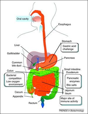
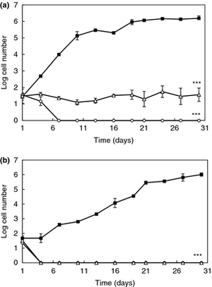
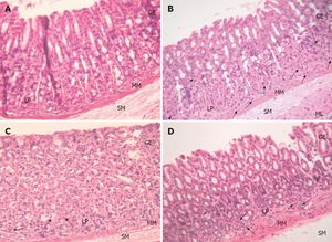
<br>[[Image:LPCrowley.png|thumb|300px|right|a) Effect of 16 and 62 cCFS on the growth of Rhodotorula mucilaginosa in orange juice. Control juice (), juice containing cCFS from Lactobacillus plantarum 16 (), juice containing cCFS from Lact. plantarum 62 (). P < 0·001 (***). (b) Effect of 16 and 62 cCFS on the growth of R. mucilaginosa in yoghurt. Control yoghurt (), yoghurt containing cCFS from Lact. plantarum 16 (), yoghurt containing cCFS from Lact. plantarum 62 (). P < 0·001 (***). Crowley 2012.]][[Image:LPSunanliganon.jpg|thumb|300px|right|Hematoxylin-eosin stained gastric sections (×20). A: Control group showed normal gastric histopathology; B: Helicobacter pylori infected group showed infiltration of inflammatory cells (arrows); C and D: Lactobacillus plantarum (L. plantarum) B7 106 CFUs/mL treated and L. plantarum B7 1010 CFUs/mL treated groups showed improvements in gastric inflammation. GE: Gastric epithelium; LP: Lamina propria; MM: Muscularis mucosae; SM: Submucosa; ML: Muscularis.Sunanliganon 2012.]]Include some current research, with at least one figure showing data.<br> | <br>[[Image:LPDaniel.jpeg|thumb|300px|right|Schematic representation of the human gastrointestinal tract (GI tract) indicating its different regions where recombinant lactic acid bacteria (rLAB) encounter various challenges. Daniel 2011.]][[Image:LPCrowley.png|thumb|300px|right|a) Effect of 16 and 62 cCFS on the growth of Rhodotorula mucilaginosa in orange juice. Control juice (), juice containing cCFS from Lactobacillus plantarum 16 (), juice containing cCFS from Lact. plantarum 62 (). P < 0·001 (***). (b) Effect of 16 and 62 cCFS on the growth of R. mucilaginosa in yoghurt. Control yoghurt (), yoghurt containing cCFS from Lact. plantarum 16 (), yoghurt containing cCFS from Lact. plantarum 62 (). P < 0·001 (***). Crowley 2012.]][[Image:LPSunanliganon.jpg|thumb|300px|right|Hematoxylin-eosin stained gastric sections (×20). A: Control group showed normal gastric histopathology; B: Helicobacter pylori infected group showed infiltration of inflammatory cells (arrows); C and D: Lactobacillus plantarum (L. plantarum) B7 106 CFUs/mL treated and L. plantarum B7 1010 CFUs/mL treated groups showed improvements in gastric inflammation. GE: Gastric epithelium; LP: Lamina propria; MM: Muscularis mucosae; SM: Submucosa; ML: Muscularis.Sunanliganon 2012.]]Include some current research, with at least one figure showing data.<br> | ||
==Conclusion== | ==Conclusion== | ||
Revision as of 18:54, 22 April 2013
Introduction
By [Student Name]
At right is a sample image insertion. It works for any image uploaded anywhere to MicrobeWiki. The insertion code consists of:
Double brackets: [[
Filename: PHIL_1181_lores.jpg
Thumbnail status: |thumb|
Pixel size: |300px|
Placement on page: |right|
Legend/credit: Electron micrograph of the Ebola Zaire virus. This was the first photo ever taken of the virus, on 10/13/1976. By Dr. F.A. Murphy, now at U.C. Davis, then at the CDC.
Closed double brackets: ]]
Other examples:
Bold
Italic
Subscript: H2O
Superscript: Fe3+
Introduce the topic of your paper. What microorganisms are of interest? Habitat? Applications for medicine and/or environment?
Section 1
Include some current research, with at least one figure showing data.
Section 2
Include some current research, with at least one figure showing data.
Section 3
Include some current research, with at least one figure showing data.
Conclusion
Overall text length at least 3,000 words, with at least 3 figures.
References
Edited by student of Joan Slonczewski for BIOL 238 Microbiology, 2011, Kenyon College.
