Murine Leukemia Virus (MuLV): Difference between revisions
From MicrobeWiki, the student-edited microbiology resource
No edit summary |
No edit summary |
||
| Line 1: | Line 1: | ||
by Rodney S. Tucker II | by Rodney S. Tucker II | ||
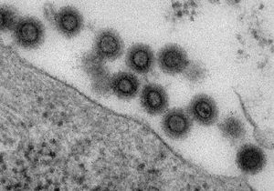
[[Image: MuLV_EM_image.jpg|thumb|300px|right|TEM of the Murine Leukemia Virus released from an infected cell. By Goff of Columbia University Medical Center.]] | [[Image: MuLV_EM_image.jpg|thumb|300px|right|TEM of the Murine Leukemia Virus released from an infected cell. By Goff of Columbia University Medical Center.]] | ||
[[Image: MuLV genome.jpg|thumb|300px|right|Murine Leukemia Virus Genome. By Alan Rein.]] | [[Image: MuLV genome.jpg|thumb|300px|right|Murine Leukemia Virus Genome. By Alan Rein.]] | ||
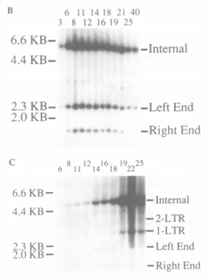
[[Image: Hirt_procedure_results.PNG|thumb|right|Hirt procedure results from Janet Hartley's study of MuLV expression in RAW264.7 cell lines.]] | |||
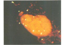
[[Image: Fluorescence_in_situ_hybridization.PNG|thumb|left|Fluorescence in situ hybridization showing the prescence of viral DNA in the nucleus.]] | |||
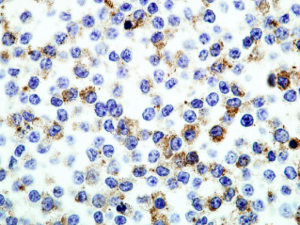
[[Image: MuLV_p30_expression.PNG|thumb|right|The expression of MuLV p30 in RAW264.7 cells. By Janet Hartley.]] | |||
Revision as of 00:27, 23 April 2013
by Rodney S. Tucker II


