File:Fruit Fly Gut.jpeg: Difference between revisions
From MicrobeWiki, the student-edited microbiology resource
Mary.cameron (talk | contribs) (A photomicrographic optical section through the tip of a ''D. melanogaster'' larval gut. The green shows activity of the Notch signaling pathway; the blue and red represent stained nuclear and cytoskeletal markers respectively. The image was captured b...) |
(No difference)
|
Latest revision as of 17:57, 1 May 2013
A photomicrographic optical section through the tip of a D. melanogaster larval gut. The green shows activity of the Notch signaling pathway; the blue and red represent stained nuclear and cytoskeletal markers respectively. The image was captured by Jessica Von Stetina of Whitehead Institute for Biomedical Research in Cambridge, Massachusetts, USA, and shared by www.nikonsmallworld.com.
File history
Click on a date/time to view the file as it appeared at that time.
| Date/Time | Thumbnail | Dimensions | User | Comment | |
|---|---|---|---|---|---|
| current | 17:57, 1 May 2013 | 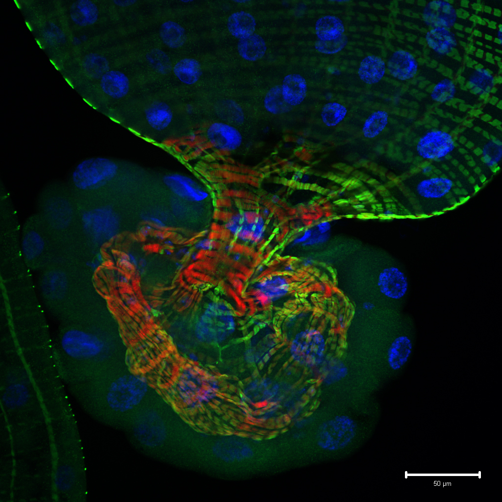 | 1,024 × 1,024 (924 KB) | Mary.cameron (talk | contribs) | A photomicrographic optical section through the tip of a ''D. melanogaster'' larval gut. The green shows activity of the Notch signaling pathway; the blue and red represent stained nuclear and cytoskeletal markers respectively. The image was captured b... |
You cannot overwrite this file.
File usage
The following page uses this file:
