File:P jejuni.jpg: Difference between revisions
From MicrobeWiki, the student-edited microbiology resource
(Scanning electron micrographs showing cells of P. jejuni, (a) strains CD3 : 27, (b) CD3 : 28T and (c) P. histicola strain CD3 : 32. (d) Transmission electron micrograph of a cell of strain CD3 : 3.) |
(No difference)
|
Latest revision as of 22:03, 12 March 2014
Scanning electron micrographs showing cells of P. jejuni, (a) strains CD3 : 27, (b) CD3 : 28T and (c) P. histicola strain CD3 : 32. (d) Transmission electron micrograph of a cell of strain CD3 : 3.
File history
Click on a date/time to view the file as it appeared at that time.
| Date/Time | Thumbnail | Dimensions | User | Comment | |
|---|---|---|---|---|---|
| current | 22:03, 12 March 2014 | 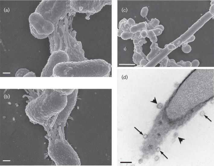 | 709 × 549 (159 KB) | Marisa.crommett (talk | contribs) | Scanning electron micrographs showing cells of P. jejuni, (a) strains CD3 : 27, (b) CD3 : 28T and (c) P. histicola strain CD3 : 32. (d) Transmission electron micrograph of a cell of strain CD3 : 3. |
You cannot overwrite this file.
File usage
The following page uses this file:
