Tannerella forsythia: Difference between revisions
No edit summary |
No edit summary |
||
| Line 1: | Line 1: | ||
{{Conway}} | |||
{{ | |||
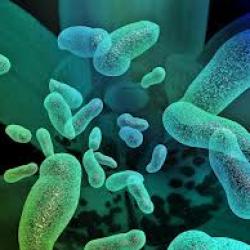
[[File:Tannerella forsythia.jpg|400px|thumb|right|Image of<i>Tannerella forsythia</i>. From: lsn.ou.edu [https://lsn.osu.edu/sites/lsn.osu.edu/files/imagesCAWMLKG2.jpg?1392648663]]] | [[File:Tannerella forsythia.jpg|400px|thumb|right|Image of<i>Tannerella forsythia</i>. From: lsn.ou.edu [https://lsn.osu.edu/sites/lsn.osu.edu/files/imagesCAWMLKG2.jpg?1392648663]]] | ||
Latest revision as of 18:55, 11 February 2016


Etiology/Bacteriology
Taxonomy
| Domain = Bacteria
| Phylum = Bacteroidetes
| Class = Bacteroidia
| Order = Bacteroidiales
| Family = Porphyromonadaceae
| Genus = Tannerella
| species = T. forsythia
Description
Tannerella forsythia is a gram-negative anaerobe previously referred to as Bacteroides forsythus until it was reclassified into a new genus :Tannerella. [1] Because of the difficulty in cultivating this pathogen until recently, much is to be learned about it, although there is overwhelming evidence in its role in the pathogenesis of periodontal disease [2][3]. Tannerella forsythia has been shown to be a periodontal pathogen because it is associated with increased levels of periodontitis, there is evidence of a host response to its antigens, it will cause disease in animal models, and because of its virulence factors which have the ability to cause disease. Periodontal disease is shown to be both a result of the direct effects of bacterial virulence factors as well as the self-damaging host immune response to the bacteria[3][8].
Pathogenesis
Epidemiology
Periodontal diseases have been recognized and treated for the last 5 millennia from writings found in ancient Egyptian and Chinese writings. The discovery of the idea that bacteria contribute to this disease is attributed to Von Leeuwenhoek, a 17th century scientist, who observed “tiny animalcules” around the teeth and attributed them to causing disease. Classification of the causes of this disease, however, has been a different issue as it’s a very multivariable illness. It was not until recently (1999) that the AAP finally settled on a complete encyclopedic classification systems of this disease that accounts for various factors such as age, sex, systemic disease, other infections, and more[4].
United States
A recent study completed by the Centers for Disease Control and Prevention (CDC) has found that approximately 50% of American Adults over the age of 30 has periodontal disease to some varying form of severity. In adults over the age of 65, is over 70%. Periodontal disease has a higher prevalence in men than in women, and highest in the Mexican American population. There is also a relation between socioeconomic status and periodontal disease with lower income being related to a worsening periodontal health status. Periodontal disease has been called one of the most prevalent non-communicable chronic diseases in the population[5].
Worldwide
Severe periodontal disease affects 10-15% of adults over the age of 30 worldwide[7]. According to WHO (World Health Organization, it is one of two worst dental diseases that affects the world [4].
Virulence factors
Trypsin-like protease and PrtH protease
Tannerella forsythia is an organism that is incapable of breaking down carbohydrates for energy (asacchrolytic) and therefore requires peptides for growth. The trypsin-like protease and the PrtH protease has been identified as essential enzymes for degrading host proteins as well as contributors for bacterial virulence due to their ability to degrade host periodontal tissues and cleaving components of the host immune response. The trypsin-like protease is mainly involved in the degradation of host proteins into smaller peptides and probably does not really play a role in virulence. The PrtH protease has the ability to cleave larger protein substrates as well as to function as a detachment factor (causing adherent cells to detach from the substratum) and subsequently release the chemokine IL-8[3]. A substantial form of this protein is called FDF(forsythia detachment factor). It functions in cell detachment and is implicated in the disintegration of sub-gingival tissue, although much remains to be elucidated on the mechanisms of how FDF interferes with cellular adhesion[9].
BspA protein
BspA is a protein that is both secreted and existing on the cell surface. Studies show that this protein binds to extracellular components fibronectin and fibrinogen and so is necessary for adherence and invasion into host epithelial cells. It also plays a role in inflammation by inducing the release of pro-inflammatory cytokines from monocytes and chemokines from epithelial cells. This cytokine secretion requires the activation of toll-like receptor 2(TLR2) in host cells and this protein seems to be the ligand that binds to this receptor[1]. In addition, this protein has been shown to mediate bacterial adherence and invasion into gingival epithelial cells[3].
Glycosidases
Tannerella forsythia has been shown to express a variety of glycosidases that play a variety of roles in its pathogenesis, including SiaHI and NanH sialidases[7]. These glycosidases theoretically play a role in the degradation of oligosaccharides and proteoglycans that would invariable affect the integrity of the periodontal cavity and tissue. This degradation can also expose protein epitopes for adherence and colonization by the bacterium itself or other potentially pathogenic bacteria. This glycosidic activity also yields to the accumulation of a toxic methylglyoxal product in vitro, which is theorized to contribute to tissue damage in hosts affected by periodontitis and colonized by this pathogen[3].
S-layer
S-layer promotes adhesion for bacterial cells allowing Tannerella forsythia to be able to adhere and invade the epithelium of the oral cavity with greater ease[3]. In addition to the S-layer mediating adhesion and/or invasion of the human gingival epithelial cells, it also is implicated in the potential to delay the recognition of Tannerella forsythia by the innate immune system of the host[8]. The surface of this pathogen also contains lipoproteins that activate host cells by the release of proinflammatory cytokines as well as inducing cellular apoptosis[3]. Additionally flagella and pilus-like structures have been discovered on this pathogen (despite it's ability to be motile) and are therefore hypothesized to play a role in pathogenesis[8].
Clinical features
Symptoms
Tannerella forsythia is one of the pathogenic species that exist in the oral cavity of the mouth and can cause periodontal disease. This disease results from an excessive host immune response or virulence factors released by pathogenic species in the oral cavity[2]. Periodontitis is a multi-factorial and variable illness that can either be acute or chronic. Ultimately, it can result in the disintegration of the tooth supporting tissues such as periodontal ligaments and alveolar bone[9]. The risk of this disease is exacerbated with poor dental hygiene, external influences, or systemic diseases such as diabetes[2].Tannerella forsythia needs N-acetylmuric acid (a vital component of the bacterial cell wall of this pathogen), which it does not produce on its own, but is rather produced by its pathogenic counterparts Porphyromonas gingivalis and Tannerella denticola. As a result its pathogenicity is a dependent[1].It’s presence in the oral cavity, however, is usually a marker for destructive periodontal disease[2].
Diagnosis
The clinical diagnosis of periodontal disease is complicated as periodontal disease encompasses a wide spectrum of diseases in itself. A healthy periodontum is stippled, and a coral pink (slightly darker or lighter pigmentation depending on race) with a knife edge margin where the gingiva meets the tooth. A departure from this concept is where disease may be classified[4]. A diagnosis of periodontitis is given to those who have obvious gingival changes that lend themselves to characteristics of gingivitis, a loss of tissue in a periodontal pocket, or other attachment loss problems[4].
Treatment
Treatment for periodontal disease is targeted at removing the pathogenic bacteria, reversing/controlling risk factors, and the prevention of colonization of the same pathogenic species[2].
SRP
This treatment is known as scaling and root planning (SRP), and involves meticulously removing/reducing the bacteria and toxins contained in a periodontal pocket. This method is not entirely effective, because for one it is very difficult to remove all bacteria and debris from the pocket, but those that are more virulent and remain in deeper parts of the pocket can rapidly recolonize the pocket[2].
Systemic and Local Treatment Adjuncts
Local antibiotic or antimicrobial treatment has been found to positively impact periodontal therapy outcomes[2].
System therapy-host modulation
Host modulation is a redirection of an inflammatory host response. The drug doxycycline is used to prevent college breakdown and has been shown to reduce pocket depth and reduce the presence of inflammatory mediators[2].
Antimicrobials
Antibiotics used for treatment include metronidazole, amoxicillin, and tetracycline. The disadvantage for antibiotic therapy is the high dosage required to battle periodontal disease. The high usages can also contribute to the increasing levels of antibiotic resistance[2].
Local Therapy
Locally administered antibiotics are effective at a lower dose and have not been implicated in antibiotic resistance. Different treatments using this method include ten percent doxycycline hyclate applied as a gel directly to pockets, minocycline hydrochloride applied via syringe directly into the pocket, or chlorhexidine gluconate[2].
Prevention
For most cases of periodontal disease, prevention is as easy as normal dental hygiene by brushing your teeth twice a day and cleaning between your teeth. However, if proper dental hygiene is abandoned, then plaque can build up and gums can be irritated and inflamed. Pathogens such as Tannerella forsythia can move into pockets that occur when the irritated gums separate from the teeth and can further irritate the gums. The pathogens then further promote irritation, leading to gingivitis and eventually periodontal disease[6]. Systemic disease such as diabetes, already promote an inflamed environment in cells and as a result it is easier for Tannerella forsythia to gain access to pockets and promote periodontal disease due to the hyperglycemic environment that the cells are exposed to. As a result, controlling the systemic disease has shown promise in controlling the periodontal disease progression[3].
Host Immune Response
These bacteria and the products that they secrete are recognized by the host innate immune response as being foreign by recognition of certain PAMP’s by PRR's of the innate immune system. As a result, chemokines and cytokines are released into the cellular environment, which results in recruitment of phagocytic cells such as neutrophils as well as a release of antimicrobial peptides. The inflammation cascade characteristic of this response can get out of hand and result in tissue destruction and increased periodontal pocket depth. In extreme cases, if the bacteria are not cleared, destruction of tooth-supporting tissue, and bone loss can result in the oral cavity[1].
References
1. Homma, K. 2009. Tannerella forsythia. A New Intruder of the Human Periodontal Pocket!. Spotlight on a lab. [Online]. [<http://wings.buffalo.edu/ubscientist/Kiyo_Epub09.pdf>]
2. Serio, Francis G. and Duncan, Teresa B. September 2009. The Pathogenesis and Treatment of Periodontal Disease. [Online].
[<http://www.ineedce.com/courses/1686/pdf/pathogenesisandtreatment.pdf>]
3. Sharma, Ashu. October 2010. Virulence Mechanisms of Tannerella forsythia.Periodontal 2000. [Online].
[<http://www.ncbi.nlm.nih.gov/pmc/articles/PMC2934765/#!po=18.3333>]
4. Highfield, J. 2009. Diagnosis and classification of periodontal disease. Australian Dental Journal. [Online].
[<http://www.onlinelibrary.wiley.com/store/10.1111/j.1834-7819.2009.01140.x/asset/j.1834-7819.2009.01140.x.pdf;jsessionid=11B663A455274539A4900009634CFB65.f02t04?v-1&t=hy4atbrn&s=204ac43598f107a6f7fa81e373377df2cf3164ac>]
5. Eke, P.I. August 2012. Prevalence of Periodontitis in Adults in the United States. American Academy of Periodontology. [Online].
[<http://www.perio.org/consumer/cdc-study.htm>]
6. American Dental Association.2001. Preventing periodontal disease. [Online].
[<http://www.ada.org/~/media/ADA/Publications/Files/patient_08.ashx>]
7. Petersen, P., Ogawa, H. 2005. Strengthening the Prevention of Periodontal Disease: The WHO approach. [Online] [<http://www.who.int/oral_health/publications/jop2005_76_12_2187.pdf?ua=1>]
8. Sekot, G. 2012. Anaylsis of the cell surface layer ultrastructure of the oral pathogen Tannerella forsythia. Arch Microbiol. [Online] [<http://download.springer.com.ezproxy.lib.ou.edu/static/pdf/187/art%253A10.1007%252Fs00203-012-0792-3.pdf?auth66=1406649745_ba3c0d15298137a75cd9e726c8cad310&ext=.pdf>]
9. Nakajima, T. 2006. Isolation and indentification of a cytopathic activity in Tannerella forsythia. Biochemical and Biophysical Research Communications. Elsevier. [Online] [<http://www.sciencedirect.com.ezproxy.lib.ou.edu/science/article/pii/S0006291X06022327>]
10. Periodontal (Gum) Disease: Causes, symptoms, and Treatments. August 2012. National Institute of Dental and Cranofacial Research. [Online] [<http://www.nidcr.nih.gov/OralHealth/Topics/GumDiseases/PeriodontalGumDisease.htm>]
Created by {Krishna Manohar}, student of Tyrrell Conway at the University of Oklahoma.
