Addition of Telomerase to Somatic Cells: Difference between revisions
(Created page with "==Introduction== Select a topic about genetics or evolution in a specific organism or ecosystem.<br> The topic must include one section about microbes (bacteria, viruses, fung...") |
|||
| (105 intermediate revisions by the same user not shown) | |||
| Line 1: | Line 1: | ||
[[image:Img-research-Telomerase_Genomics-illiustration.png|thumb|300px|right| Figure 1: Chromosome, telomere and telomerase shown together. The image was made by the Chopra Foundation https://www.choprafoundation.org/education-research/past-studies/seduction-of-spirit-meditation-study/]] | |||
Telomerase is an enzyme that is able to regenerate telomeres. In humans, it is found in some tissues, such as male germ cells, activated lymphocytes, and certain types of stem cell populations.<ref>https://science.sciencemag.org/content/266/5193/2011</ref><ref>https://onlinelibrary.wiley.com/doi/abs/10.1002/%28SICI%291520-6408%281996%2918%3A2%3C173%3A%3AAID-DVG10%3E3.0.CO%3B2-3</ref> If present in somatic cells, it can turn them cancerous. If absent, telomere shortening results which can be one of the contributing factors to aging. Hence telomerase is of the utmost importance in the telomere theory of aging as well as the theory of senescence as a tumour suppression mechanism. | |||
==Microbiome== | |||
< | Telomerase is a reverse transcriptase enzyme with its own RNA template. This makes it very similar to retroviruses with the notable exception of capsids. Endogenous retroviruses (ERVs) are endogenous viral elements in the genome that closely resemble and can be derived from retroviruses. They are abundant in the genomes of jawed vertebrates, and they comprise up to 5–8% of the human genome (lower estimates of ~1%).<ref>https://www.pnas.org/content/101/14/4894</ref><ref>https://onlinelibrary.wiley.com/doi/full/10.1111/j.1365-2249.2004.02592.x</ref> Such similarities could be used to establish the idea that viruses may have fused with the cells of other organisms to give rise to telomerase in the course of evolution from the RNA world. <ref>https://www.frontiersin.org/articles/10.3389/fonc.2012.00201/full</ref> | ||
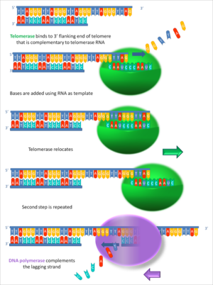
[[ | [[image:Working_principle_of_telomerase.png|thumb|300px|right| Figure 2: An image illustrating how telomerase elongates telomere ends progressively.]] | ||
==Telomeres== | |||
< | Telomeres are repeating sequences of nucleotides at the end of chromosomes which do not code for any proteins. For vertebrates, this sequence has been identified to be "TTAGGC"<ref>https://www.sciencedirect.com/science/article/pii/S009286740081908X?via%3Dihub</ref><ref>http://genesdev.cshlp.org/content/11/21/2801</ref>. Now, DNA polymerase is not capable of replicating the chromosome along its entire length. Hence every time DNA replications are undergone, the ends of chromosomes will be left out. Telomeres are extra sequences at those very ends so they can be lost instead of other vital protein-coding sequences. Leonard Hayflick demonstrated that a normal human fetal cell population will divide between 40 and 60 times in cell culture before entering a senescence phase. It occurs because each time a cell undergoes mitosis, the telomeres on the ends of each chromosome shorten slightly. Cell division will cease once telomeres shorten to a critical length. Hayflick interpreted his discovery to be aging at the cellular level.<ref>https://www.sciencedirect.com/science/article/pii/0014482761901926?via%3Dihub</ref> | ||
< | |||
==Telomerase Structure in Humans== | |||
The molecular composition of the human telomerase complex was determined by Scott Cohen and his team at the Children's Medical Research Institute (Sydney Australia) and consists of two molecules each of human telomerase reverse transcriptase (TERT), telomerase RNA (TR or TERC), and dyskerin (DKC1).<ref>https://science.sciencemag.org/content/315/5820/1850</ref> That was the whole complex but the parts of key interest: one is the functional RNA component, hTERC which serves as a template for telomeric DNA synthesis: the other is a catalytic protein, hTERT with reverse transcriptase activity <ref>https://www.ncbi.nlm.nih.gov/pubmed/9389643/</ref><ref>https://www.ncbi.nlm.nih.gov/pubmed/9328464/</ref><ref>https://www.ncbi.nlm.nih.gov/pubmed/9110970/</ref><ref>https://www.ncbi.nlm.nih.gov/pubmed/9288757/</ref>.The genes of telomerase, TERT,<ref>https://www.genenames.org/index.html</ref> TERC,<ref>https://www.genenames.org/data/hgnc_data.php?hgnc_id=11727</ref> DKC1<ref>https://www.genenames.org/data/hgnc_data.php?hgnc_id=2890</ref> and TEP1,<ref>https://www.genenames.org/data/hgnc_data.php?hgnc_id=11726</ref> are located on different chromosomes. The human TERT gene (hTERT) of roughly 4 kb, is translated into a protein that is made up of 1132 amino acids.<ref>https://www.ncbi.nlm.nih.gov/protein/109633031</ref> TERT polypeptide folds with (and carries) TERC, a non-coding RNA (451 nucleotides long). | |||
==Telomerase Gene in Human Somatic Cells== | |||
Among the core components of human telomerase, only the catalytic component hTERT seems to be the limiting determinant of telomerase activity, as other components undergo constitutive regulation. Substantial experimental data show that the transcriptional regulation of hTERT gene expression makes up the primary and rate-limiting step in the activation of telomerase to add repeats of "TTAGG".<ref>https://academic.oup.com/hmg/article/8/1/137/2356225</ref><ref>https://www.ncbi.nlm.nih.gov/pubmed/10029071/</ref><ref>https://www.sciencedirect.com/science/article/pii/S0092867400805383?via%3Dihub</ref><ref>https://www.ncbi.nlm.nih.gov/pubmed/9973199/</ref> Thus, it has been proposed that the lack of telomerase activity in most somatic human cells is due to transcriptional repression of the hTERT gene. Consistent with this hypothesis, cell fusions between normal somatic cells and some telomerase-positive immortal cells result in repression of telomerase activity <ref>https://www.sciencedirect.com/science/article/pii/S0047637499000548?via%3Dihub</ref><ref>https://www.ncbi.nlm.nih.gov/pmc/articles/PMC450086/</ref>. Importantly, microcell-mediated transfer of specific human chromosomes into cancer cells results in repression of hTERT expression and downregulation of telomerase activity <ref>https://www.sciencedirect.com/science/article/pii/S0959804997000907?via%3Dihub</ref>. These observations indicate that normal cells can always be producing functional transcriptional repressors of hTERT <ref>https://academic.oup.com/jnci/article/91/1/4/2549265</ref>. | |||
< | ==Telomerase Activity in Human Somatic Cells== | ||
Most types of normal human somatic cells, however, are telomerase negative and hence have a limited ability to duplicate themselves.<ref>https://www.sciencedirect.com/science/article/pii/0014482765902119?via%3Dihub</ref><ref>https://www.nature.com/articles/35036093</ref>. When this limit is reached, which depends on cell type and origin, cells will permanently stop dividing. This stage in the life of a cell is called its senescence stage. It has been suggested that senescence could function as a tumor suppressor mechanism to prevent the accumulation of multiple oncogenic mutations <ref>https://ehp.niehs.nih.gov/doi/10.1289/ehp.919359</ref><ref>https://www.sciencedirect.com/science/article/pii/S0959437X00001635?via%3Dihub</ref>. Telomerase activity was found to be absent in most normal human somatic cells but present in over 90% of cancerous cells and in vitro-immortalized cells <ref>https://science.sciencemag.org/content/266/5193/2011</ref><ref>https://www.sciencedirect.com/science/article/pii/S0959804997000622?via%3Dihub</ref> Hence loss of TERT repressors can be thought to result in the upregulation of hTERT expression and telomerase activity as seen during multistage carcinogenesis. | |||
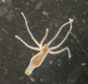
[[image:Hydravulgaris.jpg|thumb|300px|right| Figure 3: A hydra vulgaris with 6 tentacles.]] | |||
==Telomerase Activity in Other Species== | |||
<ref> | The presence of the enzyme telomerase reverse transcriptase (TERT) can stop the telomere shortening which theoretically leads to unlimited replicative capacity. On the other hand, continuous telomere-shortening might limit the ability to repair damaged cells, which, according to the telomere theory of aging, is believed to be the main reason why organisms are weakened in later age and finally show an age-related increase in mortality. However, in the nonaging Hydra species, telomerase activity lacks enough understanding. Indirect proof of a possible telomerase activity in Hydra and a prerequisite for constant cell number growth and regeneration has been deduced from the presence of a gene-encoding TERT, as identified in the genome of H. magnipapillata. <ref>https://www.sciencedirect.com/science/article/pii/S0168952510002088?via%3Dihub</ref> Other species characterized by a high level of increase in cell number, such as sponges, maintain the vitality of their telomere through a high telomerase activity. <ref>https://www.pnas.org/content/109/11/4209</ref> Many invertebrates, fish, amphibians, and reptiles that show never-ending cell number growth to maintain a high regeneration ability require high telomerase activities <ref>https://link.springer.com/chapter/10.1007%2F978-90-481-3465-6_11</ref>. These observations suggest that the clonal lineages of Hydra, with a stable cell proliferation rate, might preserve their long-term telomere integrity via telomerase activity. | ||
==Conclusion== | ==Conclusion== | ||
Telomeres are undoubtedly essential resources in the process of DNA replication. Telomerase has the potential to increase the supply of that resource infinitely. It is therefore imperative to study telomerase much more. This will require an approach that involves the studies of the structure in various species. This is to prove or disprove its impact on the aging of organisms. If telomerase is to be ever used in any anti-aging therapy then it would require further insight into its relationship with cancer otherwise it would be counterintuitive. <i>Hydra vulgaris </i> can be considered to be non-aging and hence they could be used as a model organism to study the phenomenon of telomerase activity. Manipulation of the regulation of hTERT gene may hold the key to curing cancer or senescence or both. | |||
==References== | ==References== | ||
Latest revision as of 03:30, 7 December 2019

Telomerase is an enzyme that is able to regenerate telomeres. In humans, it is found in some tissues, such as male germ cells, activated lymphocytes, and certain types of stem cell populations.[1][2] If present in somatic cells, it can turn them cancerous. If absent, telomere shortening results which can be one of the contributing factors to aging. Hence telomerase is of the utmost importance in the telomere theory of aging as well as the theory of senescence as a tumour suppression mechanism.
Microbiome
Telomerase is a reverse transcriptase enzyme with its own RNA template. This makes it very similar to retroviruses with the notable exception of capsids. Endogenous retroviruses (ERVs) are endogenous viral elements in the genome that closely resemble and can be derived from retroviruses. They are abundant in the genomes of jawed vertebrates, and they comprise up to 5–8% of the human genome (lower estimates of ~1%).[3][4] Such similarities could be used to establish the idea that viruses may have fused with the cells of other organisms to give rise to telomerase in the course of evolution from the RNA world. [5]
Telomeres
Telomeres are repeating sequences of nucleotides at the end of chromosomes which do not code for any proteins. For vertebrates, this sequence has been identified to be "TTAGGC"[6][7]. Now, DNA polymerase is not capable of replicating the chromosome along its entire length. Hence every time DNA replications are undergone, the ends of chromosomes will be left out. Telomeres are extra sequences at those very ends so they can be lost instead of other vital protein-coding sequences. Leonard Hayflick demonstrated that a normal human fetal cell population will divide between 40 and 60 times in cell culture before entering a senescence phase. It occurs because each time a cell undergoes mitosis, the telomeres on the ends of each chromosome shorten slightly. Cell division will cease once telomeres shorten to a critical length. Hayflick interpreted his discovery to be aging at the cellular level.[8]
Telomerase Structure in Humans
The molecular composition of the human telomerase complex was determined by Scott Cohen and his team at the Children's Medical Research Institute (Sydney Australia) and consists of two molecules each of human telomerase reverse transcriptase (TERT), telomerase RNA (TR or TERC), and dyskerin (DKC1).[9] That was the whole complex but the parts of key interest: one is the functional RNA component, hTERC which serves as a template for telomeric DNA synthesis: the other is a catalytic protein, hTERT with reverse transcriptase activity [10][11][12][13].The genes of telomerase, TERT,[14] TERC,[15] DKC1[16] and TEP1,[17] are located on different chromosomes. The human TERT gene (hTERT) of roughly 4 kb, is translated into a protein that is made up of 1132 amino acids.[18] TERT polypeptide folds with (and carries) TERC, a non-coding RNA (451 nucleotides long).
Telomerase Gene in Human Somatic Cells
Among the core components of human telomerase, only the catalytic component hTERT seems to be the limiting determinant of telomerase activity, as other components undergo constitutive regulation. Substantial experimental data show that the transcriptional regulation of hTERT gene expression makes up the primary and rate-limiting step in the activation of telomerase to add repeats of "TTAGG".[19][20][21][22] Thus, it has been proposed that the lack of telomerase activity in most somatic human cells is due to transcriptional repression of the hTERT gene. Consistent with this hypothesis, cell fusions between normal somatic cells and some telomerase-positive immortal cells result in repression of telomerase activity [23][24]. Importantly, microcell-mediated transfer of specific human chromosomes into cancer cells results in repression of hTERT expression and downregulation of telomerase activity [25]. These observations indicate that normal cells can always be producing functional transcriptional repressors of hTERT [26].
Telomerase Activity in Human Somatic Cells
Most types of normal human somatic cells, however, are telomerase negative and hence have a limited ability to duplicate themselves.[27][28]. When this limit is reached, which depends on cell type and origin, cells will permanently stop dividing. This stage in the life of a cell is called its senescence stage. It has been suggested that senescence could function as a tumor suppressor mechanism to prevent the accumulation of multiple oncogenic mutations [29][30]. Telomerase activity was found to be absent in most normal human somatic cells but present in over 90% of cancerous cells and in vitro-immortalized cells [31][32] Hence loss of TERT repressors can be thought to result in the upregulation of hTERT expression and telomerase activity as seen during multistage carcinogenesis.
Telomerase Activity in Other Species
The presence of the enzyme telomerase reverse transcriptase (TERT) can stop the telomere shortening which theoretically leads to unlimited replicative capacity. On the other hand, continuous telomere-shortening might limit the ability to repair damaged cells, which, according to the telomere theory of aging, is believed to be the main reason why organisms are weakened in later age and finally show an age-related increase in mortality. However, in the nonaging Hydra species, telomerase activity lacks enough understanding. Indirect proof of a possible telomerase activity in Hydra and a prerequisite for constant cell number growth and regeneration has been deduced from the presence of a gene-encoding TERT, as identified in the genome of H. magnipapillata. [33] Other species characterized by a high level of increase in cell number, such as sponges, maintain the vitality of their telomere through a high telomerase activity. [34] Many invertebrates, fish, amphibians, and reptiles that show never-ending cell number growth to maintain a high regeneration ability require high telomerase activities [35]. These observations suggest that the clonal lineages of Hydra, with a stable cell proliferation rate, might preserve their long-term telomere integrity via telomerase activity.
Conclusion
Telomeres are undoubtedly essential resources in the process of DNA replication. Telomerase has the potential to increase the supply of that resource infinitely. It is therefore imperative to study telomerase much more. This will require an approach that involves the studies of the structure in various species. This is to prove or disprove its impact on the aging of organisms. If telomerase is to be ever used in any anti-aging therapy then it would require further insight into its relationship with cancer otherwise it would be counterintuitive. Hydra vulgaris can be considered to be non-aging and hence they could be used as a model organism to study the phenomenon of telomerase activity. Manipulation of the regulation of hTERT gene may hold the key to curing cancer or senescence or both.
References
- ↑ https://science.sciencemag.org/content/266/5193/2011
- ↑ https://onlinelibrary.wiley.com/doi/abs/10.1002/%28SICI%291520-6408%281996%2918%3A2%3C173%3A%3AAID-DVG10%3E3.0.CO%3B2-3
- ↑ https://www.pnas.org/content/101/14/4894
- ↑ https://onlinelibrary.wiley.com/doi/full/10.1111/j.1365-2249.2004.02592.x
- ↑ https://www.frontiersin.org/articles/10.3389/fonc.2012.00201/full
- ↑ https://www.sciencedirect.com/science/article/pii/S009286740081908X?via%3Dihub
- ↑ http://genesdev.cshlp.org/content/11/21/2801
- ↑ https://www.sciencedirect.com/science/article/pii/0014482761901926?via%3Dihub
- ↑ https://science.sciencemag.org/content/315/5820/1850
- ↑ https://www.ncbi.nlm.nih.gov/pubmed/9389643/
- ↑ https://www.ncbi.nlm.nih.gov/pubmed/9328464/
- ↑ https://www.ncbi.nlm.nih.gov/pubmed/9110970/
- ↑ https://www.ncbi.nlm.nih.gov/pubmed/9288757/
- ↑ https://www.genenames.org/index.html
- ↑ https://www.genenames.org/data/hgnc_data.php?hgnc_id=11727
- ↑ https://www.genenames.org/data/hgnc_data.php?hgnc_id=2890
- ↑ https://www.genenames.org/data/hgnc_data.php?hgnc_id=11726
- ↑ https://www.ncbi.nlm.nih.gov/protein/109633031
- ↑ https://academic.oup.com/hmg/article/8/1/137/2356225
- ↑ https://www.ncbi.nlm.nih.gov/pubmed/10029071/
- ↑ https://www.sciencedirect.com/science/article/pii/S0092867400805383?via%3Dihub
- ↑ https://www.ncbi.nlm.nih.gov/pubmed/9973199/
- ↑ https://www.sciencedirect.com/science/article/pii/S0047637499000548?via%3Dihub
- ↑ https://www.ncbi.nlm.nih.gov/pmc/articles/PMC450086/
- ↑ https://www.sciencedirect.com/science/article/pii/S0959804997000907?via%3Dihub
- ↑ https://academic.oup.com/jnci/article/91/1/4/2549265
- ↑ https://www.sciencedirect.com/science/article/pii/0014482765902119?via%3Dihub
- ↑ https://www.nature.com/articles/35036093
- ↑ https://ehp.niehs.nih.gov/doi/10.1289/ehp.919359
- ↑ https://www.sciencedirect.com/science/article/pii/S0959437X00001635?via%3Dihub
- ↑ https://science.sciencemag.org/content/266/5193/2011
- ↑ https://www.sciencedirect.com/science/article/pii/S0959804997000622?via%3Dihub
- ↑ https://www.sciencedirect.com/science/article/pii/S0168952510002088?via%3Dihub
- ↑ https://www.pnas.org/content/109/11/4209
- ↑ https://link.springer.com/chapter/10.1007%2F978-90-481-3465-6_11
Edited by Nafeez Ishmam Ahmed, student of Joan Slonczewski for BIOL 116 Information in Living Systems, 2019, Kenyon College.


