Citrobacter koseri: Difference between revisions
No edit summary |
No edit summary |
||
| (32 intermediate revisions by the same user not shown) | |||
| Line 1: | Line 1: | ||
{{Uncurated}} | {{Uncurated}} | ||
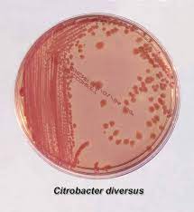
[ | [[Image:citrobacter-koseri_fromberg_drainvocht_04_f-350x220 (2).jpg|thumb|300px|right| Growth on a petri dish of the bacteria, <i >Citrobacter koseri</i >. Elmicrobiologist.]] | ||
==Classification== | ==Classification== | ||
| Line 16: | Line 16: | ||
|} | |} | ||
<i >Citrobacter koseri</i > | |||
==Description and Significance== | ==Description and Significance== | ||
<i>Citrobacter koseri<i> (formerly Citrobacter diversus)is a member of the Enterobacteriaceae. It has the appearance of straight rods and occurs singly in pairs. The length of the bacillus is 1 micrometer in diameter by 2.0-6.0 micrometers in length. <i>Citrobacter koseri<i> is found in soil, water, and in the intestinal tracts of both humans and animals. It has a negative importance in regards to the cause of neonatal meningitis and brain abscess formation. | <i >Citrobacter koseri</i > (formerly <i >Citrobacter diversus</i >)is a member of the Enterobacteriaceae. It has the appearance of straight rods and occurs singly in pairs. The length of the bacillus is 1 micrometer in diameter by 2.0-6.0 micrometers in length. <i >Citrobacter koseri</i > is found in soil, water, and in the intestinal tracts of both humans and animals. It has a negative importance in regards to the cause of neonatal meningitis and brain abscess formation. | ||
==Genome Structure== | ==Genome Structure== | ||
The genome size varies between 4.0 to 5.0 Mb. The GC content of the genomes is between 51 to 56%. It is unknown in the amount of chromosomes it has. C. koseri is circular. The genome consist of 4,536 protein-coding sequences. | The genome size varies between 4.0 to 5.0 Mb. The GC content of the genomes is between 51 to 56%. It is unknown in the amount of chromosomes it has. <i >C. koseri</i > is circular. The genome consist of 4,536 protein-coding sequences. | ||
==Cell Structure, Metabolism and Life Cycle== | ==Cell Structure, Metabolism and Life Cycle== | ||
<i>Citrobacter koseri<i> gains energy solely through using citrate as a carbon source. The molecule it was shown to produce when isolated was hemolysin, which is a substance found in the blood that kills red blood cells and free hemoglobin. | <i >Citrobacter koseri</i > gains energy solely through using citrate as a carbon source. The molecule it was shown to produce when isolated was hemolysin, which is a substance found in the blood that kills red blood cells and free hemoglobin. | ||
==Ecology and Pathogenesis== | ==Ecology and Pathogenesis== | ||
<i>Citrobacter koseri<i> inhabitants the intestinal tracts of animals and humans. It does not benefit or work together with another organism. <i>C. koseri<i> is known to be linked with hospital-acquired infections. It does not positively contribute to environment but rather | <i >Citrobacter koseri</i > inhabitants the intestinal tracts of animals and humans. It does not benefit or work together with another organism. <i >C. koseri</i > is known to be linked with hospital-acquired infections. It does not positively contribute to environment but rather causes contamination of an area where it inhabits. | ||
<i >Citrobacter koseri</i >causes urinary tract infections. Animals and humans are the targets for host. The virulence factors are connected with flagellar apparatus biosynthesis and iron uptake. Symptoms of the microbe include sepsis, meningitis, and cerebritis, seizures, apnea, becoming sickly, and a bulging fontanelle. | |||
The image below shows two cow brains, one affected by <i >Citrobacter koseri</i > and one unaffected. The brain to the left shows a calf that suffered from meningitis from the microbe, <i >Citrobacter koseri</i >. The affected brain displays cloudiness and thickening of the meninges. | |||
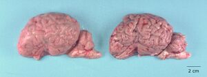
[[Image:microb.jpg|300px|center|<i >Citrobacter koseri</i > Septicaemia in a Holstein Calf. M. Komine, A. Massa, L. Moon & T. Mullaney.]] | |||
==References== | ==References== | ||
[ | [https://www.sciencedirect.com/science/article/pii/B9781455748013002204. Michael S.Donnenberg, and Michael S.Donnenberg. “Enterobacteriaceae.” Mandell, Douglas, and Bennett's Principles and Practice of Infectious Diseases (Eighth Edition), W.B. Saunders, 31 Oct. 2014, .] | ||
Fecteau et al., 2009 | |||
G. Fecteau, B.P. Smith, L.W. George | |||
Septicemia and meningitis in the newborn calf | |||
Veterinary Clinics of North America: Food Animal Practice, 25 (2009), pp. 195-208 | |||
[https://www.sciencedirect.com/science/article/pii/B9781416040446501175. G.Fisher, Randall. “Citrobacter.” Feigin and Cherry's Textbook of Pediatric Infectious Diseases (Sixth Edition), W.B. Saunders, 21 Mar. 2012,] | |||
[https://www.sciencedirect.com/science/article/pii/B9780128053515000065. AudreyWangerVioletaChavezRichard S.P.HuangAmerWahedJeffrey K.ActorAmitavaDasgupta, et al. “Overview of Bacteria.” Microbiology and Molecular Diagnosis in Pathology, Elsevier, 23 June 2017.] | |||
[https://www.frontiersin.org/articles/10.3389/fmicb.2019.02774/full Yuan, C., Yin, Z., Wang, J., Qian, C., Wei, Y., Zhang, S., Jiang, L., & Liu, B. (1AD, January 1). Comparative genomic analysis of Citrobacter and key genes essential for the pathogenicity of Citrobacter Koseri. Frontiers.] | [https://www.frontiersin.org/articles/10.3389/fmicb.2019.02774/full Yuan, C., Yin, Z., Wang, J., Qian, C., Wei, Y., Zhang, S., Jiang, L., & Liu, B. (1AD, January 1). Comparative genomic analysis of Citrobacter and key genes essential for the pathogenicity of Citrobacter Koseri. Frontiers.] | ||
[https://www.ncbi.nlm.nih.gov/pmc/articles/PMC7729419/ Ohkubo, T., Matsumoto, Y., Cho, O., Ogasawara, Y., & Sugita, T. (2020, December 10). Complete genome sequence of Citrobacter Koseri strain MPUCK001. Microbiology resource announcements.] | [https://www.ncbi.nlm.nih.gov/pmc/articles/PMC7729419/ Ohkubo, T., Matsumoto, Y., Cho, O., Ogasawara, Y., & Sugita, T. (2020, December 10). Complete genome sequence of Citrobacter Koseri strain MPUCK001. Microbiology resource announcements.] | ||
==Author== | ==Author== | ||
Latest revision as of 23:29, 12 December 2022
Classification
Bacteria; Proteobacteria; Gammaproteobacteria; Enterobacterales; Enterobacteriaceae
Species
|
NCBI: [1] |
Citrobacter koseri
Description and Significance
Citrobacter koseri (formerly Citrobacter diversus)is a member of the Enterobacteriaceae. It has the appearance of straight rods and occurs singly in pairs. The length of the bacillus is 1 micrometer in diameter by 2.0-6.0 micrometers in length. Citrobacter koseri is found in soil, water, and in the intestinal tracts of both humans and animals. It has a negative importance in regards to the cause of neonatal meningitis and brain abscess formation.
Genome Structure
The genome size varies between 4.0 to 5.0 Mb. The GC content of the genomes is between 51 to 56%. It is unknown in the amount of chromosomes it has. C. koseri is circular. The genome consist of 4,536 protein-coding sequences.
Cell Structure, Metabolism and Life Cycle
Citrobacter koseri gains energy solely through using citrate as a carbon source. The molecule it was shown to produce when isolated was hemolysin, which is a substance found in the blood that kills red blood cells and free hemoglobin.
Ecology and Pathogenesis
Citrobacter koseri inhabitants the intestinal tracts of animals and humans. It does not benefit or work together with another organism. C. koseri is known to be linked with hospital-acquired infections. It does not positively contribute to environment but rather causes contamination of an area where it inhabits.
Citrobacter kosericauses urinary tract infections. Animals and humans are the targets for host. The virulence factors are connected with flagellar apparatus biosynthesis and iron uptake. Symptoms of the microbe include sepsis, meningitis, and cerebritis, seizures, apnea, becoming sickly, and a bulging fontanelle.
The image below shows two cow brains, one affected by Citrobacter koseri and one unaffected. The brain to the left shows a calf that suffered from meningitis from the microbe, Citrobacter koseri. The affected brain displays cloudiness and thickening of the meninges.
References
Fecteau et al., 2009 G. Fecteau, B.P. Smith, L.W. George Septicemia and meningitis in the newborn calf Veterinary Clinics of North America: Food Animal Practice, 25 (2009), pp. 195-208
Author
Page authored by Kassidy Cartret, student of Prof. Bradley Tolar at UNC Wilmington.


