Tectiviridae: Difference between revisions
(→Author) |
|||
| (2 intermediate revisions by the same user not shown) | |||
| Line 30: | Line 30: | ||
[[File:Structure..jpeg|frameless|caption]] | [[File:Structure..jpeg|frameless|caption]] | ||
This figure represents the genome, more specifically highlighting that there are more promoters (6) than operons (5) which makes our virus unique! | |||
==Cell Structure, Metabolism and Life Cycle== | ==Cell Structure, Metabolism and Life Cycle== | ||
Viruses like Alphatectivirus PRD1 do not have their own metabolism and are non-living entities that are unable to generate ATP and translate it to form proteins. Instead, Alphatectivirus PRD1 relies on the metabolism of host cells to provide energy and metabolic substances for their life cycles. It is icosahedral in shape, has no external envelope but does have a capsid along with spike proteins (Raoult). It also has an inner membrane vesicle enclosed by the capsid that is made up of virus encoded proteins and lipids from the host cells plasma membrane (Tuma 2021). | Viruses like Alphatectivirus PRD1 do not have their own metabolism and are non-living entities that are unable to generate ATP and translate it to form proteins. Instead, Alphatectivirus PRD1 relies on the metabolism of host cells to provide energy and metabolic substances for their life cycles. It is icosahedral in shape, has no external envelope but does have a capsid along with spike proteins (Raoult). It also has an inner membrane vesicle enclosed by the capsid that is made up of virus encoded proteins and lipids from the host cells plasma membrane (Tuma 2021). | ||
| Line 47: | Line 46: | ||
In terms of symbiosis, PRD1 is parasitic to bacteria, but it also promotes horizontal gene transfer, which helps increase genetic diversity among bacterial populations (Parra 2024). By facilitating the exchange of genetic material, PRD1 indirectly supports microbial adaptability and evolution. The virus's activity can also contribute to biogeochemical cycles by lysing bacterial cells, which releases nutrients such as carbon and nitrogen back into the environment (Parra 2024). These nutrients fuel microbial activity, promoting the growth of other organisms and contributing to the recycling of essential elements. | In terms of symbiosis, PRD1 is parasitic to bacteria, but it also promotes horizontal gene transfer, which helps increase genetic diversity among bacterial populations (Parra 2024). By facilitating the exchange of genetic material, PRD1 indirectly supports microbial adaptability and evolution. The virus's activity can also contribute to biogeochemical cycles by lysing bacterial cells, which releases nutrients such as carbon and nitrogen back into the environment (Parra 2024). These nutrients fuel microbial activity, promoting the growth of other organisms and contributing to the recycling of essential elements. | ||
PRD1 does not cause disease in humans, animals, or plants, as it specifically targets bacteria (Parra 2024). Its "virulence" is limited to its interaction with bacterial hosts, where it uses unique viral mechanisms to penetrate and degrade the bacterial cell wall. This interaction results in bacterial lysis, which can influence microbial populations but does not lead to disease in higher organisms. PRD1’s role is more ecological, helping to maintain microbial diversity and supporting environmental nutrient cycles. | PRD1 does not cause disease in humans, animals, or plants, as it specifically targets bacteria (Parra 2024). Its "virulence" is limited to its interaction with bacterial hosts, where it uses unique viral mechanisms to penetrate and degrade the bacterial cell wall. This interaction results in bacterial lysis, which can influence microbial populations but does not lead to disease in higher organisms. PRD1’s role is more ecological, helping to maintain microbial diversity and supporting environmental nutrient cycles. More specifically our microbe could be used in biotechnology in hospital wastewater. PRD1 could be used to reduce bacterial loads and prevent the spread of harmful pathogens from hospital wastewater into the environment (Parra 2024). This approach could be a greener alternative to chemical disinfectants, which may harm ecosystems. | ||
==References== | ==References== | ||
Latest revision as of 03:20, 4 December 2024
Classification
Viruses; Varidnaviria; Bamfordvirae; Preplasmiviricota; Tectiliviricetes; Kalamavirales; Tectiviridae; Alphatectivirus; Alphatectivirus PRD1
Species
NCBI: [1]
Enterobacteria phage PRD1
Description and Significance
Alphatectivirus PRD1 is a bacteriophage known for its unique structural features. PRD1 has an icosahedral capsid, measuring about 66 nanometers in diameter, and unlike many bacteriophages, it possesses an internal lipid membrane situated beneath its protein shell (Raoult). This rare characteristic among icosahedral viruses contributes to PRD1’s stability in diverse environments, including extreme pH and temperature conditions. Its linear double-stranded DNA genome, spanning approximately 15,000 base pairs, encodes multiple structural proteins that are crucial for the assembly and function of both the capsid and the internal membrane.
PRD1's primary habitat involves environments where Gram-negative bacteria, such as Escherichia coli and Salmonella, thrive, which includes various soil and water ecosystems, as well as potential niches in the intestines of animals (Wilhelm 1999). This phage’s resilience across environmental conditions highlights its potential as a candidate for studies in bacteriophage therapy, particularly against antibiotic-resistant bacterial strains. PRD1’s bacteriophage characteristics allow it to target specific bacteria, raising the possibility of developing bacteriophage-based treatments that could serve as an alternative to conventional antibiotics.
In addition to its relevance in combating bacterial infections, PRD1 has proven valuable in studies of DNA replication, protein synthesis, and virus-host interactions ("UniProt Fall Back Message"). Its relatively simple genome and replication mechanisms make it an ideal model for understanding virus assembly and gene regulation. Furthermore, PRD1’s unique lipid-containing capsid has inspired advancements in nanotechnology and drug delivery research, where scientists aim to mimic its stability and efficiency in viral assembly for use in biotechnological applications (Stanton 2024).
Genome Structure
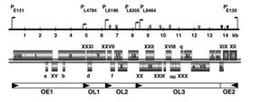
Single molecule of linear dsDNA that is 14927 bp, its entire sequence is known ("Tectiviridae (Taxid:10656)"). The 5' ends of the DNA have covalently linked proteins. It also has inverted terminal repeats at both ends of the DNA sequence which serve as origins of replication (Nehaus 2024). There are 31 genes in the genome but only 26 of them have a known protein product, the other 5 have listed under product "hypothetical protein". It also has 5 operons and 6 promoters, the extra promoter is due to the fact that it can be replicated from both ends of the DNA sequence due to the inverted terminal repeats (Nehaus 2024). After infection and DNA entry the capsid proteins polymerize in the cytoplasm while the membrane associated proteins are inserted into the host's plasma membrane.
 This figure represents the genome, more specifically highlighting that there are more promoters (6) than operons (5) which makes our virus unique!
Cell Structure, Metabolism and Life Cycle
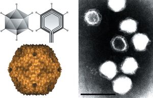
Viruses like Alphatectivirus PRD1 do not have their own metabolism and are non-living entities that are unable to generate ATP and translate it to form proteins. Instead, Alphatectivirus PRD1 relies on the metabolism of host cells to provide energy and metabolic substances for their life cycles. It is icosahedral in shape, has no external envelope but does have a capsid along with spike proteins (Raoult). It also has an inner membrane vesicle enclosed by the capsid that is made up of virus encoded proteins and lipids from the host cells plasma membrane (Tuma 2021).
The life cycle of Tectiviridae viruses begins with adsorption, which is where the virion attaches to the host cells surface (Caruso 2019). Following absorption, the virion extrudes a tube structure through a vertex to deliver its genome into the host cell (Caruso 2019). Then the host cell will replicate of the viral genome, through the DNA strand displacement, while transcription is achieved via DNA-templated processes (Caruso 2019). As the viral genome is replicated and transcribed, capsid proteins polymerize around a lipoprotein vesicle, which is translocated within the cytoplasm by virion assembly factors (Tuma 2021). Finally, mature virions are released through lysis of the host cell (Caruso 2019). This process of cell lysis in Tectiviridae, such as in the case of PRD1, is facilitated by virus-encoded lysis machinery, including four key proteins: P15 (endolysin), P35 (holin), P36, and P37.
Tectiviridae phages are unique because they can form a special structure that is partly made of proteins and partly made of lipids. This structure acts like a protective bubble around the virus particles. When it's time for the new viruses to leave the host cell, this bubble can help them interact with the cell's membrane. When the virus disrupts the host cells membrane it causes the host cell to lyse. On the other hand, the virus can fuse with the host cells membrane to create a pathway for the virus to leave the host cell without destroying it. In either case, the bubble helps the viruses escape and spread to infect new cells.
Ecology and Pathogenesis
Alphatectivirus PRD1 is a bacteriophage that infects Gram-negative bacteria, including species like Pseudomonas putida and Escherichia coli. It inhabits environments with dense bacterial populations, such as soil, water, and wastewater, where it contributes to microbial community dynamics by regulating bacterial populations. The virus relies on its bacterial hosts for replication, and in turn, it can influence ecological balance by controlling the density of specific bacterial species ("Tectiviridae (Taxid:10656)"). It does not infect humans, animals, or plants, but is integral to microbial processes in these environments (Harvey, 2004).
In terms of symbiosis, PRD1 is parasitic to bacteria, but it also promotes horizontal gene transfer, which helps increase genetic diversity among bacterial populations (Parra 2024). By facilitating the exchange of genetic material, PRD1 indirectly supports microbial adaptability and evolution. The virus's activity can also contribute to biogeochemical cycles by lysing bacterial cells, which releases nutrients such as carbon and nitrogen back into the environment (Parra 2024). These nutrients fuel microbial activity, promoting the growth of other organisms and contributing to the recycling of essential elements.
PRD1 does not cause disease in humans, animals, or plants, as it specifically targets bacteria (Parra 2024). Its "virulence" is limited to its interaction with bacterial hosts, where it uses unique viral mechanisms to penetrate and degrade the bacterial cell wall. This interaction results in bacterial lysis, which can influence microbial populations but does not lead to disease in higher organisms. PRD1’s role is more ecological, helping to maintain microbial diversity and supporting environmental nutrient cycles. More specifically our microbe could be used in biotechnology in hospital wastewater. PRD1 could be used to reduce bacterial loads and prevent the spread of harmful pathogens from hospital wastewater into the environment (Parra 2024). This approach could be a greener alternative to chemical disinfectants, which may harm ecosystems.
References
Caruso, Steven M., et al. “A Novel Genus of Actinobacterial Tectiviridae.” MDPI, Multidisciplinary Digital Publishing Institute, 7 Dec. 2019, www.mdpi.com/1999-4915/11/12/1134.
Neuhaus, Alyssa. “Viral Vectors 101: Inverted Terminal Repeats.” Addgene Blog, blog.addgene.org/viral-vectors-101-inverted-terminal-repeats. Accessed 27 Nov. 2024.
Parra, Boris, et al. “Characterization and Abundance of Plasmid-Dependent Alphatectivirus Bacteriophages.” Microbial Ecology, U.S. National Library of Medicine, 27 June 2024, pmc.ncbi.nlm.nih.gov/articles/PMC11211187/.
Raoult, D. “Enterobacteria Phage PRD1.” Enterobacteria Phage PRD1 - an Overview | ScienceDirect Topics, www.sciencedirect.com/topics/immunology-and-microbiology/enterobacteria-phage-prd1#:~:text=The%20capsid%20of%20Enterobacteria%20phage,are%20used%20for%20receptor%20recognition. Accessed 27 Nov. 2024.
Stanton, Cassandra R., et al. “Isolation of a PRD1-like Phage Uncovers the Carriage of Three Putative Conjugative Plasmids in Clinical Burkholderia Contaminans.” Research in Microbiology, Elsevier Masson, 4 Apr. 2024, www.sciencedirect.com/science/article/pii/S0923250824000330.
Sokolova, A., et al. “Solution Structure of Bacteriophage PRD1 Vertex Complex.” The Journal of Biological Chemistry, U.S. National Library of Medicine, pubmed.ncbi.nlm.nih.gov/11577098. Accessed 27 Nov. 2024.
“Tectiviridae (Taxid:10656).” ViralZone, viralzone.expasy.org/160?outline=all_by_species. Accessed 27 Nov. 2024.
Tuma, Roman, et al. “Membrane-Containing Icosahedral DNA Bacteriophages.” Encyclopedia of Virology (Fourth Edition), Academic Press, 1 Mar. 2021, www.sciencedirect.com/science/article/pii/B9780128145159000448.
“Uniprot Website Fallback Message.” UniProt, www.uniprot.org/uniprotkb/P09009/entry. Accessed 27 Nov. 2024.
Wilhelm, Steven W., and Curtis A. Suttle. “Viruses and Nutrient Cycles in the Sea: Viruses Play Critical Roles in the Structure and Function of Aquatic Food Webs.” OUP Academic, Oxford University Press, 1 Oct. 1999, academic.oup.com/bioscience/article/49/10/781/222807?searchresult=1.
Author
Page authored by Abi Miller, Lee Hinson, Mariella Dagdag, & Alexis Grimes, students of Prof. Bradley Tolar at UNC Wilmington.

