Cysticercosis: Difference between revisions
| Line 38: | Line 38: | ||
==Clinical features== | ==Clinical features== | ||
Cysticercosis is an infectious disease that causes the development of cysts, called cysticerci, in various locations of the body (7). The most popular locations for the cysticerci are the subcutaneous tissues, skeletal muscle, eyes, brain, and spinal cord. The location of the cysts determines a lot about the symptoms, severity, and treatment of the disease. (8) | |||
===Symptoms=== | ===Symptoms=== | ||
There are a variety of symptoms that can be demonstrated depending on the location and number of the cysts. If they are located in the skeletal muscle or subcutaneous tissue, there will often times be no signs of symptoms (7). However, it is sometimes possible to feel lumps and palpable nodules beneath the skin, which may become tender and cause localized pain. If the cysts are located in the eye (usually in the subretinal space or vitreous humor), vision might become blurry and disturbed. This is due to inflammation or retinal detachment. (8) Cysts in the brain or spinal cord are cause a specific type of cysticercosis called neurocysticercosis. This is the most severe type of this infectious disease and causes the most threatening symptoms. Symptoms of neurocysticercosis include: headache, seizures, confusion, difficulty with balance, gait disturbances, brain swelling, hydrocephalus (excess fluid around the brain), stroke, and even death (7). In fact, cysticercosis is the single most common cause of acquired epileptic seizures (3). Obviously, the symptoms of this disease vary depending on the location of the cysts. | |||
===Morbidity and Mortality=== | ===Morbidity and Mortality=== | ||
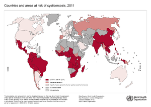
[[File:Global distribution.png|300px|thumb|right|Global distribution of cysticercosis. From: WHO [http://www.who.int/taeniasis/Global_distribution_cysticercosis_2011.png?ua=1]]] | |||
Cysticercosis is the most common parasitic disease across the world, and it is estimated that 50 million people are infected with it. The highest rates of infection for cysticercosis are found in parts of Africa, Asia, Mexico, and Central and South America. (8) However, it is becoming more of an issue in the United States because of increased immigration and world travelling. The listed locations tend to have poor sanitation and free-roaming pigs, which are environments that <i>Taenia solium</i> thrive and fecal-oral contamination can more easily occur. (9) | |||
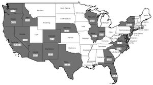
[[File:US Distribution.JPEG|300px|thumb|left|Fatal cysticercosis cases by state 1990-2002. From: NCBI [http://www.ncbi.nlm.nih.gov/pmc/articles/PMC2725874/]]] | |||
In the US, cysticercosis is considered by the CDC as one of the five Neglected Parasitic Infections (NPIs). This means that cysticercosis has been targeted as a priority for public health action. (9) From 1990-2002 in the United States, there were 221 cysticercosis deaths. Most of the people who died were Latino, and more were men than women. Nearly 85% of the people who died were foreign born, and 62% had emigrated from Mexico. California accounted for 57% of the total deaths. Only 33 of the 221 deaths occurred in US-born persons. (10) | |||
Neurocysticercosis is the most common parasitic disease of the nervous system worldwide. It is estimated that more than 1,000 new cases are diagnosed each year in the United States alone. (8) Also, the WHO estimates that of the 50 million people in the world who are affected by epilepsy, 80% of them live in low-income and lower-middle-income countries where <i>T. solium</i> is an endemic (11). | |||
==Diagnosis== | ==Diagnosis== | ||
Revision as of 23:07, 23 July 2014


Etiology/Bacteriology
Taxonomy
| Domain = Eukaryota | Phylum = Platyhelminthes | Class = Cestoda | Order = Cyclophyllidea | Family = Taeniidae | Genus = Taenia | species = T. solium
Description
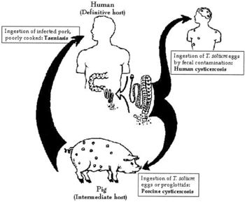
Cysticercosis is an infectious disease that is caused by the larvae of Taenia solium. T. solium is a two-host zoonotic cestode whose adult stage consists of a 2-4 meter long tapeworm. The only known final host for this adult stage is humans, specifically in the small intestine. The intermediate host is the pig, so part of the T. solium life cycle takes place in that host prior to causing taeniasis in humans. Taeniasis is a parasitic infection caused by the presence of tapeworms in the intestine. A person with taeniasis is capable of infecting themselves or others with cysticercosis through fecal-oral transmission. Cysticercosis is transmitted by eating food or drinking water that is contaminated with T. solium eggs. 2-3 months after ingestion, the eggs develop into cysticerci, which are cysts formed by T. solium larvae. The location of the cysts throughout the body determines the symptoms that are demonstrated, diagnoses performed, and treatment used. If the cysts are located in the central nervous system, the infection is specifically called neurocysticercosis.
Cysticercosis is most prevalent in parts of Africa, Asia, Mexico, and Central and South America. However, it is a worldwide issue with about 50 million people having the infection. Because of its fecal-oral transmission, it is important to prevent consumption of contaminated food and water and practice proper hand washing techniques. If the parasite does infect a host, the host immune response will attempt to remove the infection through innate and adaptive responses. Inflammation is a key to the host immune response, yet it can hurt the host as much as it benefits it.
Pathogenesis
Transmission
Larvae of the tapeworm Taenia solium are what cause cysticercosis. If a person becomes contaminated with an adult tapeworm, an infection called taeniasis, they can shed T. solium eggs in their stool. These eggs must then be consumed through fecal-oral route transmission, which can occur by drinking water or eating food that is contaminated with T. solium eggs, or by putting a fecal contaminated object in one’s mouth. Due to this form of transmission, it is possible for autoinfection to occur. If a person has taeniasis, they can infect themselves with the T. solium eggs they shed and thus develop cysticercosis. (12)
Infectious dose, incubation, and colonization
The infectious dose required to cause cysticercosis is relatively unknown (15). The median incubation period for cysticercosis is estimated to be about 3.5 years (13) and can range from 10 days to 10 years (14). A person who has consumed T. solium may go for years without having any symptoms of infection. This is due to the fact that cysticerci often only cause symptoms when they are dying or become large enough to interfere with vital organs. (14) Furthermore, it normally takes 2-3 months for the eggs to develop into cysticerci (15).

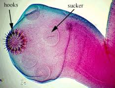
Upon consumption of T. solium eggs, the oncospheres hatch in the intestine and then penetrate the intestinal wall. T. solium contain sharp, spine hooks and four suckers that help them adhere to and penetrate the gut wall (17). They then migrate to skeletal muscle, the brain, eyes, subcutaneous tissues, and other places where they develop into cysticerci. (16)
Epidemiology
Taenia solium is prevalent in parts of Africa, Asia, Mexico, and Latin America where humans live in close proximity to pigs. This is because pigs are one of the hosts during the life cycle of this two-host parasite, while humans are the other host. When pigs eat T. solium eggs, the eggs develop into larvae and form cysts within the pig’s muscles and other tissues. Then, if humans consume undercooked or raw infected pork, they swallow these cysts. The larvae then come out of their cysts within the human gut and develop into adult tapeworms. Now, this person will shed T. solium eggs in their stool. At this point, if fecal-oral contamination occurs, cysticercosis will develop in whoever consumed the contaminated fecal matter. (12) It is clear that cysticercosis is spread by fecal-oral route, not the consumption of undercooked, infected pork meat.
Virulence factors
T. solium contains a number of virulence factors that allow it to survive amidst the host immune response. The oncospheres invade the host and are susceptible to antibody and complement responses, but by the time the host actually mounts an antibody response, the parasite begins to transform to the more resistant larval stage, called metacestode. During the larval stage, the parasite can evade complement-mediated destruction. This is achieved by a few different virulence factors including paramyosin, which is a protein that inhibits C1q; taeniaestatin, a proteinase, inhibits the classical and alternate pathways; and sulfated polysaccharides, which activate complement away from the parasite. The larval stage of T. solium also manipulates antibody production in such a way that the host produces antibody that cannot kill the mature metacestode, but instead provides a protein source to the parasite. Lastly, taeniaestatin may interfere with lymphocyte proliferation and macrophage functions; therefore, compromising the cellular immune response. (19)
Clinical features
Cysticercosis is an infectious disease that causes the development of cysts, called cysticerci, in various locations of the body (7). The most popular locations for the cysticerci are the subcutaneous tissues, skeletal muscle, eyes, brain, and spinal cord. The location of the cysts determines a lot about the symptoms, severity, and treatment of the disease. (8)
Symptoms
There are a variety of symptoms that can be demonstrated depending on the location and number of the cysts. If they are located in the skeletal muscle or subcutaneous tissue, there will often times be no signs of symptoms (7). However, it is sometimes possible to feel lumps and palpable nodules beneath the skin, which may become tender and cause localized pain. If the cysts are located in the eye (usually in the subretinal space or vitreous humor), vision might become blurry and disturbed. This is due to inflammation or retinal detachment. (8) Cysts in the brain or spinal cord are cause a specific type of cysticercosis called neurocysticercosis. This is the most severe type of this infectious disease and causes the most threatening symptoms. Symptoms of neurocysticercosis include: headache, seizures, confusion, difficulty with balance, gait disturbances, brain swelling, hydrocephalus (excess fluid around the brain), stroke, and even death (7). In fact, cysticercosis is the single most common cause of acquired epileptic seizures (3). Obviously, the symptoms of this disease vary depending on the location of the cysts.
Morbidity and Mortality

Cysticercosis is the most common parasitic disease across the world, and it is estimated that 50 million people are infected with it. The highest rates of infection for cysticercosis are found in parts of Africa, Asia, Mexico, and Central and South America. (8) However, it is becoming more of an issue in the United States because of increased immigration and world travelling. The listed locations tend to have poor sanitation and free-roaming pigs, which are environments that Taenia solium thrive and fecal-oral contamination can more easily occur. (9)

In the US, cysticercosis is considered by the CDC as one of the five Neglected Parasitic Infections (NPIs). This means that cysticercosis has been targeted as a priority for public health action. (9) From 1990-2002 in the United States, there were 221 cysticercosis deaths. Most of the people who died were Latino, and more were men than women. Nearly 85% of the people who died were foreign born, and 62% had emigrated from Mexico. California accounted for 57% of the total deaths. Only 33 of the 221 deaths occurred in US-born persons. (10)
Neurocysticercosis is the most common parasitic disease of the nervous system worldwide. It is estimated that more than 1,000 new cases are diagnosed each year in the United States alone. (8) Also, the WHO estimates that of the 50 million people in the world who are affected by epilepsy, 80% of them live in low-income and lower-middle-income countries where T. solium is an endemic (11).
Diagnosis
Treatment
Prevention
Host Immune Response
References
References
Created by {Jordan Voth}, students of Tyrrell Conway at the University of Oklahoma.
