Chloroquine Resistance in Plasmodium falciparum: Difference between revisions
| Line 29: | Line 29: | ||
==Mechanism of Chloroquine resistance== | ==Mechanism of Chloroquine resistance== | ||
Within the last few decades, Chloroquine was considered the best and more widely used antimalarial drug due to its effectiveness and its reasonable cost. Unfortunately, the P. falciparum parasite developed resistance to chloroquine. These resistance was mostly observed in malaria-endemic countries.<br> | Within the last few decades, Chloroquine was considered the best and more widely used antimalarial drug due to its effectiveness and its reasonable cost. Unfortunately, the <br>P. falciparum<br> parasite developed resistance to chloroquine. These resistance was mostly observed in malaria-endemic countries.<br><br> | ||
<br> | |||
In its Erythrocyte stage, P. falciparum invades the white blood cells where it forms a lysosomal isolated compartment known as the digestive vacuole (DV). The parasite grows in the erythrocite by ingesting haemoglobin from the host cell cytosol and depositing it in the digestive vacuole. The protein is degraded in the DV to its component peptides and hemewhich is incorporated into the inert and harmless crystalline polymer hemozoin(1). Chloroquine is a diprotic weak base and at low physiological pH (~7.4), it can be found in its unprotonated form, CQ, monoprotonated from, CQ+ and diprotonated , CQ++ forms. The unchanrged chloroquine is membrane permeable and it freely diffuses into the erythrocyte and into the DV. Inside the DV, Chloroquine becomes protonated. Since the membrane is not permeable to any charged species, the chloroquine accumulates in the DV. Charged chloroquine is believed to bind to haematin, a toxic byproduct of the haemoglobin proteolysis which prevents its incorporation into the haemozoin crystal. Free haematin seems to interfere with the parasite detoxificationprocess therefore damaging the plasmodium membrane(1). | |||
Chloroquine sensitive parasites accumulate more chloroquine in their DV than chloroquine resistant strains. Reduced chloroquine accumulation in the resistant parasite strains have been associated with the mutation in the gene encoding for plasmodium falciparum chloroquine resistant promoter (PfCRT). PfCRT is localized in the digestive vacuole and it is believed to aid in the exiting of positively charged chloroquine which decreases the concentration of chloroquine in the DV. It is however not clear if PfCRT acts as a carrier or a channel. The recent finding however suggests that PfCRT is an active carrier belonging to the family of Drug/Metabolite transporter superfamily (1). | |||
==References== | ==References== | ||
Revision as of 04:08, 15 April 2015
Introduction

By Edna Kemboi
At right is a sample image insertion. It works for any image uploaded anywhere to MicrobeWiki. The insertion code consists of:
Double brackets: [[
Filename: PHIL_1181_lores.jpg
Thumbnail status: |thumb|
Pixel size: |300px|
Placement on page: |right|
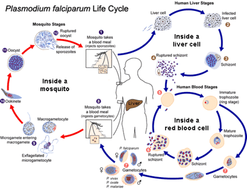
Legend/credit: Plasmodium falciparum life cycle [1].
Closed double brackets: ]]
Other examples:
Bold
Italic
Subscript: H2O
Superscript: Fe3+
Introduce the topic of your paper. What microorganisms are of interest? Habitat? Applications for medicine and/or environment?
Section 1
Include some current research, with at least one figure showing data.
Plasmodium falciparum life cycle

Include some current research, with at least one figure showing data.
Mechanism of Chloroquine resistance
Within the last few decades, Chloroquine was considered the best and more widely used antimalarial drug due to its effectiveness and its reasonable cost. Unfortunately, the
P. falciparum
parasite developed resistance to chloroquine. These resistance was mostly observed in malaria-endemic countries.
In its Erythrocyte stage, P. falciparum invades the white blood cells where it forms a lysosomal isolated compartment known as the digestive vacuole (DV). The parasite grows in the erythrocite by ingesting haemoglobin from the host cell cytosol and depositing it in the digestive vacuole. The protein is degraded in the DV to its component peptides and hemewhich is incorporated into the inert and harmless crystalline polymer hemozoin(1). Chloroquine is a diprotic weak base and at low physiological pH (~7.4), it can be found in its unprotonated form, CQ, monoprotonated from, CQ+ and diprotonated , CQ++ forms. The unchanrged chloroquine is membrane permeable and it freely diffuses into the erythrocyte and into the DV. Inside the DV, Chloroquine becomes protonated. Since the membrane is not permeable to any charged species, the chloroquine accumulates in the DV. Charged chloroquine is believed to bind to haematin, a toxic byproduct of the haemoglobin proteolysis which prevents its incorporation into the haemozoin crystal. Free haematin seems to interfere with the parasite detoxificationprocess therefore damaging the plasmodium membrane(1).
Chloroquine sensitive parasites accumulate more chloroquine in their DV than chloroquine resistant strains. Reduced chloroquine accumulation in the resistant parasite strains have been associated with the mutation in the gene encoding for plasmodium falciparum chloroquine resistant promoter (PfCRT). PfCRT is localized in the digestive vacuole and it is believed to aid in the exiting of positively charged chloroquine which decreases the concentration of chloroquine in the DV. It is however not clear if PfCRT acts as a carrier or a channel. The recent finding however suggests that PfCRT is an active carrier belonging to the family of Drug/Metabolite transporter superfamily (1).
References
[1] Hodgkin, J. and Partridge, F.A. "Caenorhabditis elegans meets microsporidia: the nematode killers from Paris." 2008. PLoS Biology 6:2634-2637.
Authored for BIOL 238 Microbiology, taught by Joan Slonczewski, 2015, Kenyon College.
