Dermabacter hominis: Difference between revisions
No edit summary |
No edit summary |
||
| Line 24: | Line 24: | ||
==Cell and colony structure== | ==Cell and colony structure== | ||
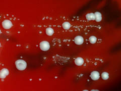
[[File:konsilium_426.jpg|thumb|DESCRIPTION]] | |||
''D. hominis'' are non-motile, mesophilic, and rod shaped. Before assignment to its current genus, ''D. hominis'' was grouped with coryneform groups 3 and 5 of the Center for Disease Control. It forms white, convex, creamy or dry colonies on nutrient and blood agar. The colonies grow to 1-1.5 millimeters in diameter after 48 hours. | ''D. hominis'' are non-motile, mesophilic, and rod shaped. Before assignment to its current genus, ''D. hominis'' was grouped with coryneform groups 3 and 5 of the Center for Disease Control. It forms white, convex, creamy or dry colonies on nutrient and blood agar. The colonies grow to 1-1.5 millimeters in diameter after 48 hours. | ||
Revision as of 17:41, 12 December 2015
A Microbial Biorealm page on the genus Dermabacter hominis
Classification
Higher order taxa
Bacteria; Actinobacteria; Actinobacteria (class); Microccocales; Dermabacteraceae
Species
|
NCBI: Taxonomy |
Dermabacter hominis
Description and significance
Dermabacter hominis can be found most commonly on human skin. It has also been isolated in a variety of other clinical specimens, including abscesses, infections of bone, a wound, or an eye, and blood cultures. Though considered an opportunistic human pathogen when first isolated in 1988, there has not been any observed direct mortality in relation to D. hominis (as of January 2014).
Genome structure
A draft genome sequence of D. hominis 1368, a multidrug resistant strain of the species D. hominis, revealed 2,507,630 base pairs. Also included in the draft genome of D. hominis 1368 are one non-coding RNA gene, 21 pseudogenes, 48 tRNA genes, and 2,227 protein-coding genes.
Cell and colony structure
D. hominis are non-motile, mesophilic, and rod shaped. Before assignment to its current genus, D. hominis was grouped with coryneform groups 3 and 5 of the Center for Disease Control. It forms white, convex, creamy or dry colonies on nutrient and blood agar. The colonies grow to 1-1.5 millimeters in diameter after 48 hours.
Metabolism
A facultative anaerobe, D. hominis is able to ferment glucose, maltose, sucrose and lactose. D. hominis is irregularly shaped (some sources list it as being coccus-shaped while others list it as rod-shaped) and is catalase positive and able to decarboxylate ornithine and lysine.
Ecology
Habitat; symbiosis; contributions to the environment. metagenomic data link
Pathology
Though there are no recorded cases of mortality as a direct result of the presence of D. hominis, it has caused peritonitis (an inflammation of the peritoneum) in a patient receiving renal replacement therapy with peritoneal dialysis.
D. hominis is resistant to daptomycin, which is rare for gram-positive organisms. As of 2014, no definitive reason for this resistance was known, though it was not thought that the resistance was due to use of daptomycin (which is a common cause of resistance).
References
[http://doi.org/10.1128/genomeA.00728-14; Albersmeier, A., Bomholt, C., Glaub, A., Rückert, C., Soriano, F., Fernández-Natal, I., & Tauch, A. (2014). Draft Genome Sequence of the Multidrug-Resistant Clinical Isolate Dermabacter hominis 1368. Genome Announcements, 2(4), e00728–14.}
[http://doi.org/10.1002/2052-2975.31; Fernández-Natal, I., Sáez-Nieto, J. A., Medina-Pascual, M. J., Albersmeier, A., Valdezate, S., Guerra-Laso, J. M., … Soriano, F. (2013). Dermabacter hominis: a usually daptomycin-resistant gram-positive organism infrequently isolated from human clinical samples. New Microbes and New Infections, 1(3), 35–40.}
[http://doi.org/10.1128/JCM.39.6.2356-2357.2001; Gómez-Garcés, J. L., Oteo, J., García, G., Aracil, B., Alós, J. I., & Funke, G. (2001). Bacteremia by Dermabacter hominis, a Rare Pathogen. Journal of Clinical Microbiology, 39(6), 2356–2357.}
[http://www.ncbi.nlm.nih.gov/pmc/articles/PMC263903/pdf/jcm00008-0096.pdf; Gruner, E., Steigerwalt, A. G., Hollis, D. G., Weyant, R. S., Weaver, R. E., Moss, C. W., … Brenner, D. J. (1994). Recognition of Dermabacter hominis, formerly CDC fermentative coryneform group 3 and group 5, as a potential human pathogen. Journal of Clinical Microbiology, 32(8), 1918–1922.} [doi:10.1099/ijs.0.028217-0.]
[http://doi.org/10.1128/JCM.39.9.3420-3421.2001; Radtke, A., Bergh, K., Øien, C. M., & Bevanger, L. S. (2001). Peritoneal Dialysis-Associated Peritonitis Caused by Dermabacter hominis. Journal of Clinical Microbiology, 39(9), 3420–3421.}
[http://www.microbiologyresearch.org/docserver/fulltext/ijsem/44/3/ijs-44-3-583.pdf?expires=1449940194&id=id&accname=guest&checksum=69677DA902D9116428BE8200DC74729C; Ca J., Collins M. Phylogenetic Analysis of Species of the meso-Diaminopimelic Acid-Containing Genera Brevibacterium and Demabacter. Int J Syst Evol Microbiol 44(3):583-585.}
Edited by W. David Nesher of Dr. Lisa R. Moore, University of Southern Maine, Department of Biological Sciences, http://www.usm.maine.edu/bio

