Sheep's wool: Difference between revisions
| Line 12: | Line 12: | ||
<br> | <br> | ||
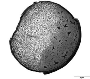
[[image: wool_cross_section.jpg | thumb | Figure 2. The cross-section of a wool fiber showing the arrangement of para- and ortho-cortical cells. <ref>Science Learning Hub – Pokapū Akoranga Pūtaiao, The University of Waikato Te Whare Wānanga o Waikato, www.sciencelearn.org.nz</ref>]] | [[image: wool_cross_section.jpg | thumb | left | Figure 2. The cross-section of a wool fiber showing the arrangement of para- and ortho-cortical cells. <ref>Science Learning Hub – Pokapū Akoranga Pūtaiao, The University of Waikato Te Whare Wānanga o Waikato, www.sciencelearn.org.nz</ref>]] | ||
Cortical cells are helix shaped and generally categorised into para- and ortho- cortical cells, with meso- cortical cells having characteristics in between the para- and ortho- categories. Para-cortical cells are loosely packed and have large amounts of cystine, while ortho-cortical cells contain discrete macrofibrils and are high in tyrosine.<ref name="marshall">Marshall RC, Orwin DF, Gillespie JM. Structure and biochemistry of mammalian hard keratin. <i>Electron Microsc Rev.</i> 1991;4(1):47-83. doi:10.1016/0892-0354(91)90016-6</ref> It was previously believed that wool’s crimp was caused by the distribution of para- and ortho-cortical cells because wool fibers show well defined bilateral segmentation in the cortex.<ref name="wortmann"></ref><ref name="marshall"></ref> However, further research has disproved the association between para and ortho-cuticle cell distribution. <ref>Hynd PI, Edwards NM, Hebart M, McDowall M, Clark S. Wool fibre crimp is determined by mitotic asymmetry and position of final keratinisation and not ortho- and para-cortical cell segmentation. Animal. 2009;3(6):838-843. doi:10.1017/S1751731109003966</ref><ref name="harland">Harland DP, Vernon JA, Woods JL, et al. Intrinsic curvature in wool fibres is determined by the relative length of orthocortical and paracortical cells. J Exp Biol. 2018;221(Pt 6):jeb172312. Published 2018 Mar 22. doi:10.1242/jeb.172312 | Cortical cells are helix shaped and generally categorised into para- and ortho- cortical cells, with meso- cortical cells having characteristics in between the para- and ortho- categories. Para-cortical cells are loosely packed and have large amounts of cystine, while ortho-cortical cells contain discrete macrofibrils and are high in tyrosine.<ref name="marshall">Marshall RC, Orwin DF, Gillespie JM. Structure and biochemistry of mammalian hard keratin. <i>Electron Microsc Rev.</i> 1991;4(1):47-83. doi:10.1016/0892-0354(91)90016-6</ref> It was previously believed that wool’s crimp was caused by the distribution of para- and ortho-cortical cells because wool fibers show well defined bilateral segmentation in the cortex.<ref name="wortmann"></ref><ref name="marshall"></ref> However, further research has disproved the association between para and ortho-cuticle cell distribution. <ref>Hynd PI, Edwards NM, Hebart M, McDowall M, Clark S. Wool fibre crimp is determined by mitotic asymmetry and position of final keratinisation and not ortho- and para-cortical cell segmentation. Animal. 2009;3(6):838-843. doi:10.1017/S1751731109003966</ref><ref name="harland">Harland DP, Vernon JA, Woods JL, et al. Intrinsic curvature in wool fibres is determined by the relative length of orthocortical and paracortical cells. J Exp Biol. 2018;221(Pt 6):jeb172312. Published 2018 Mar 22. doi:10.1242/jeb.172312 | ||
Revision as of 17:37, 11 December 2024
Introduction

Sheep (Ovis aries) have been selectively bred to continuously produce single coated wool fleece rather than coats composed of an outer hair layer and an inner wool layer.[2] True wool, as opposed to hair, is characterised by its high follicle density in the skin, small diameter, and high crimp [3]
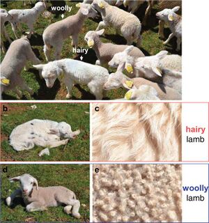
The single woolly coat is recessive trait caused by the insertion of an antisense EIF2S2 retrogene[4] into the 3′ untranslated region of the IRF2BP2 gene.[1] This gene mutation creates a chimeric IRF2BP2/asEIF2S2 RNA transcript that targets the genuine sense EIF2S2 mRNA and creates EIF2S2 dsRNA that regulates the production of EIF2S2 protein [1]. Differences in EIF2S2 expression are visible in lamb's wool: a woolly lamb has visible curls or ringlets while a hairy lamb has more of a wave pattern (Figure 1).
Wool structure and composition
All hair and wool fibers are composed of an cuticle layer of overlapping cells wrapped around a cortex. Coarse wools and many animal fibers also contain a medulla consisting of empty vacuoles.[5]
Wool has a cuticle layer that is only one cell thick, while human hair, for example, has a cuticle layer up to 10 cells thick. Wool cuticle cells also have a wedge-shaped shaped cross-section as opposed to rectangular, so the exposed edge height of wool cuticle cells is about 1 um as opposed to < 0.5 um in other animal fibers.[5]

Cortical cells are helix shaped and generally categorised into para- and ortho- cortical cells, with meso- cortical cells having characteristics in between the para- and ortho- categories. Para-cortical cells are loosely packed and have large amounts of cystine, while ortho-cortical cells contain discrete macrofibrils and are high in tyrosine.[7] It was previously believed that wool’s crimp was caused by the distribution of para- and ortho-cortical cells because wool fibers show well defined bilateral segmentation in the cortex.[5][7] However, further research has disproved the association between para and ortho-cuticle cell distribution. [8][9] Crimp is actually caused by the differences in length between para- and ortho-cuticle cells.[9]
Microbial interactions with wool
There are many microbes naturally present in sheep’s wool with >95% of bacteria found on the outer ends of the fleece and relatively few on the sheep’s skin and innermost parts of the fleece.[10]
Overpopulation of certain bacteria such as Pseudomonas aeroginosa is associated with one of wool farming’s primary concerns – blowfly infestation (also called flystrike).[11].In prolonged moisture, the protective waxy layer of sheep skin breaks down and allows P. aeroginosa and other opportunistic bacteria to multiply. This causes fleece rot – matted wool, staining or discoloration, and skin lesions. Fleece rot caused by P. aeroginosa stains the wool green.[11] Flystrike is not only encouraged by the damp conditions and vulnerable skin associated with fleece rot, the bacterial odors can attract female blowflies and stimulate oviposition. [12]
Microbial products have potential industrial applications, such as proteases being used for superwash treatment (degrading the cuticle cells so that wool can be machine washed and dried without felting)[13] This process is difficult to apply on an industrial scale and comes with concerns about the proteases damaging the entire wool fiber rather than just the cuticle. Studies[13] are being done on finding new proteases by studying microbes naturally present on wool.
Another potential application of microbial products is the use of biosurfectants to clean wool. [14]
References
- ↑ 1.0 1.1 1.2 Demars J, Cano M, Drouilhet L, et al. Genome-Wide Identification of the Mutation Underlying Fleece Variation and Discriminating Ancestral Hairy Species from Modern Woolly Sheep. Mol Biol Evol. 2017;34(7):1722-1729. doi:10.1093/molbev/msx114
- ↑ [Ryder M. A survey of European primitive breeds of sheep. Ann Genet Sel Anim. 1981;13(4):381-418. doi:10.1186/1297-9686-13-4-38]
- ↑ Doyle EK, Preston JWV, McGregor BA, Hynd PI. The science behind the wool industry. The importance and value of wool production from sheep. Anim Front. 2021;11(2):15-23. Published 2021 May 17. doi:10.1093/af/vfab005
- ↑ Staszak K, Makałowska I. Cancer, Retrogenes, and Evolution. Life (Basel). 2021;11(1):72. Published 2021 Jan 19. doi:10.3390/life11010072
- ↑ 5.0 5.1 5.2 Wortmann, F.-J. (2009). The structure and properties of wool and hair fibres. Handbook of Textile Fibre Structure, 108–145. doi:10.1533/9781845697310.1.108
- ↑ Science Learning Hub – Pokapū Akoranga Pūtaiao, The University of Waikato Te Whare Wānanga o Waikato, www.sciencelearn.org.nz
- ↑ 7.0 7.1 Marshall RC, Orwin DF, Gillespie JM. Structure and biochemistry of mammalian hard keratin. Electron Microsc Rev. 1991;4(1):47-83. doi:10.1016/0892-0354(91)90016-6
- ↑ Hynd PI, Edwards NM, Hebart M, McDowall M, Clark S. Wool fibre crimp is determined by mitotic asymmetry and position of final keratinisation and not ortho- and para-cortical cell segmentation. Animal. 2009;3(6):838-843. doi:10.1017/S1751731109003966
- ↑ 9.0 9.1 Harland DP, Vernon JA, Woods JL, et al. Intrinsic curvature in wool fibres is determined by the relative length of orthocortical and paracortical cells. J Exp Biol. 2018;221(Pt 6):jeb172312. Published 2018 Mar 22. doi:10.1242/jeb.172312
- ↑ Jackson, T. A. et al. (2002) ‘Abundance and distribution of microbial populations in sheep fleece’, New Zealand Journal of Agricultural Research, 45(1), pp. 49–55. doi: 10.1080/00288233.2002.9513492.
- ↑ 11.0 11.1 Norris BJ, Colditz IG, Dixon TJ. Fleece rot and dermatophilosis in sheep. Vet Microbiol. 2008;128(3-4):217-230. doi:10.1016/j.vetmic.2007.10.024
- ↑ Emmens RL, Murray MD. The role of bacterial odours in oviposition by Lucilia cuprina (Wiedemann) (Diptera: Calliphoridae), the Australian sheep blowfly. Bulletin of Entomological Research. 1982;72(3):367-375. doi:10.1017/S0007485300013547
- ↑ 13.0 13.1 Queiroga AC et al. (2007). Novel microbial-mediated modifications of wool. Enzyme and Microbial Technology. doi: 10.1016/j.enzmictec.2006.10.037
- ↑ Jibia SA et al. (2017) ‘Biodegradation of Wool by Bacteria and Fungi and Enhancement of Wool Quality by Biosurfactant Washing’, Journal of Natural Fibers, 15(2), pp. 287–295. doi: 10.1080/15440478.2017.1325430
Edited by Isaac Yu, student of Joan Slonczewski for BIOL 116, 2024, Kenyon College.
